
4
Review of the Scar Rule for Determining Compliance with the Horse Protection Act
This chapter reviews the scar rule, its limitations, and what changes are currently documented regarding the skin of Tennessee walking horses (TWHs) that are suspected of being sore. The chapter focuses on the evaluation of changes in the skin of the forelimb of TWHs as part of the inspection process for ensuring compliance with the “scar rule” as defined in the Horse Protection Regulations. Evaluation of these changes is an essential component of the inspection process for detection of soreness in TWHs. Particular emphasis is placed on the accuracy and specificity of the language of the scar rule in light of changes that have occurred in the TWH industry since the scar rule was included in the Horse Protection Regulations. A suggestion for updating the language of the scar rule to accurately reflect the character of soring lesions is presented. Accurate recognition and documentation of the skin abnormalities found in TWHs determined to be in violation of the scar rule is essential for training inspectors to recognize these changes and for ensuring compliance with Horse Protection Regulations. An overview of the microscopic anatomy of the skin and a review of both the current clinical abnormalities and histological (microscopic changes) of sore horses is presented and a correlation of the two is made.
THE HORSE PROTECTION ACT AND APPLICATION OF THE SCAR RULE
As discussed in Chapter 2, the Horse Protection Regulations outline the process for the examination of the forelimb of a horse before it is allowed to show and after winning in its class (post-show). The inspection of horses for compliance with the Horse Protection Act (HPA) includes a dermatologic examination of the forelimbs from below the carpus, with particular attention paid to the skin of the pastern and the coronary band. The following sections describe the specific requirements in Title 9 of the Code of Federal Regulations for these examinations:
§ 11.21(a)(2): The DQP [designated qualified person] should digitally palpate the front limbs from the knee (carpus) to the hoof with particular attention to the pastern and the fetlock. They should pick up and examine the posterior surface of the pastern and apply digital pressure to the pocket (sulcus), including the bulbs of the heel, and continue to the medial and lateral surfaces of the pastern. They should extend the foot and leg to examine [the] anterior surface including the coronary band. They may examine the rear legs after showing or any horse exhibiting lesions on or unusual movement of the rear legs. They should also inspect to determine whether the horse is scar rule compliant.
§ 11.3 Scar Rule:1 The scar rule applies to all horses born on or after October 1, 1975. Horses subject to this rule that do not meet the following scar rule criteria shall be considered to be “sore” and are subject to all prohibitions of section 5 of the Act. The scar rule criteria are as follows:
___________________
1 See https://www.law.cornell.edu/cfr/text/9/11.3 (accessed November 19, 2019).
- The anterior and anterior-lateral surfaces of the fore pasterns (extensor surface) must be free of bilateral granulomas,2 other bilateral pathological evidence of inflammation, and other bilateral evidence of abuse indicative of soring including, but not limited to, excessive loss of hair.
- The posterior surfaces of the pasterns (flexor surface), including the sulcus or “pocket” may show bilateral areas of uniformly thickened epithelial tissue if such areas are free of proliferating granuloma tissue, irritation, moisture, edema, or other evidence of inflammation.
CLINICAL DERMATOLOGIC EXAMINATION, MICROSCOPIC ANATOMY OF THE SKIN, AND PERTINENT DEFINITIONS
The dermatologic examination that is performed at the point of inspection to assess whether there is a scar rule violation is limited to the detection of gross lesions of the skin. The term “gross” refers to the clinical appearance of the skin to include what can be detected with a visual inspection by naked eye and abnormalities that can be detected by palpation and sometimes smell.
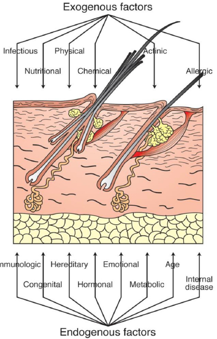
The abnormal findings documented from a dermatological examination of the skin all fall into the broad category of “lesions.” A lesion is defined as any abnormality in a tissue or organ caused by trauma or disease. As shown in Figure 4-1, many exogenous and endogenous factors can affect the integrity of the skin.
___________________
2Granuloma is defined as any one of a rather large group of fairly distinctive focal lesions that are formed as a result of inflammatory reactions caused by biological, chemical, or physical agents. (44 FR 25179, Apr. 27, 1979, as amended at 53 FR 14782, Apr. 26, 1988, 53 FR 28373, July 28, 1988.)
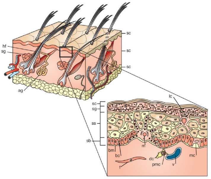
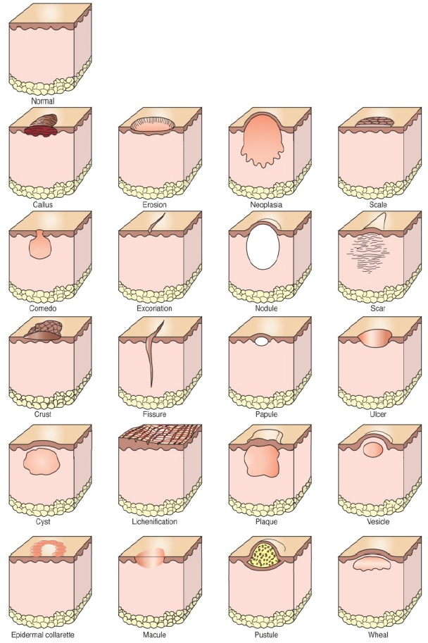
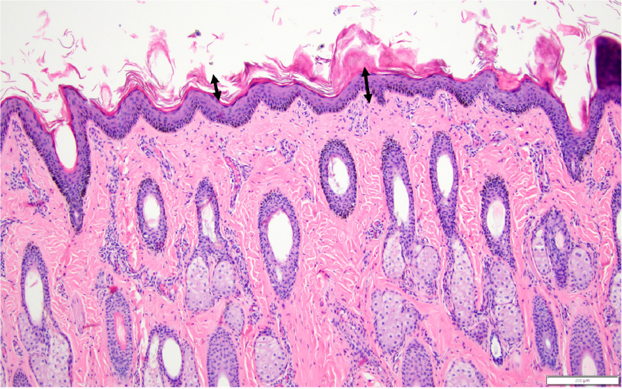
The skin has three main layers, as illustrated in Figure 4-2. Gross lesions of the skin are categorized as primary, indicating a change in a tissue that represents the effects of the original injury or disease as it first occurred. Primary lesions are the most useful lesions in determining the etiology or cause of an injury or disease. Examples of primary injuries of the skin include vesicles (blister), papules, nodules, or lacerations. Secondary lesions of the skin, on the other hand, reflect changes in the tissue that occurred over a period of time after the initial injury (Figure 4-3). Primary lesions often change in characteristic ways over time, making it possible for an experienced examiner to devise a differential list of types of initial injuries or diseases that could have produced the secondary lesion. Primary lesions are most often acute and transient, whereas secondary lesions are chronic and more persistent unless the initiating causes can be identified and removed. Figure 4-4 shows a primary (acute) lesion evolving into a secondary (chronic) lesion, which is what happens if a laceration or cut is not properly sutured or bandaged. This leads to the tissue being unable to return to its original form, resulting in the formation of the chronic and end stage of a scar. Similarly, an injury leading to a vesicle or blister over time can lead to the secondary more chronic lesion of an erosion or ulcer. In some instances, deep ulcers can also lead to scar formation over time.
A more detailed and definitive evaluation of the lesions of the skin requires the microscopic evaluation of tissue biopsies taken from the lesions. Again, primary lesions are the most useful in determining the cause of the lesions, so primary lesions are the lesion of choice for histopathological evaluation.

MICROSCOPIC EVALUATION OF SKIN BIOPSIES OF TENNESSEE WALKING HORSES FOUND TO BE IN VIOLATION OF THE SCAR RULE
To date, no peer-reviewed studies have been published on microscopic lesions of the skin from horses determined to have either skin lesions or other types of violations of the scar rule. In fact, the Horse Protection Regulations and the scar rule were written without any microscopic evaluation of skin lesions from horses suspected of being in violation of the HPA or scar rule. However, an unpublished but peer-reviewed study (Stromberg, 2017) that evaluated 136 pastern biopsies (right and left pastern from each horse) from 68 TWHs that had been disqualified for violations of the scar rule during the Celebration events of 2015 and 2016 was made available to the committee by a representative of the TWH industry. In this study, 6-mm punch biopsies were collected from the right and left palmar aspect of the pastern from each of the 68 TWHs. The skin biopsies were evaluated independently by two well-respected veterinary anatomic pathologists3 certified by the American College of Veterinary Pathologists. According to the manuscript the two pathologists agreed in their reports of abnormal findings of variable (moderate to severe) epidermal hyperplasia in the form of acanthosis (thickening of the stratum spinosum layer of the epidermis) and variable degrees of hyperkeratosis (thickening of the stratum corneum layer of the epidermis; see Figure 4-5a,b). The hyperkeratosis varied from mild to severe. Other, less consistent findings included folliculitis, follicular atrophy, and follicular distortion and mild changes in elastin fibers. The evaluators did not find any evidence of scar tissue or granulomatous inflammation and therefore concluded there was no basis or proof of scar rule violation. The selection of the appropriate site to biopsy is heavily dependent upon the recognition and understanding of the clinical (gross) lesions present. Unfortunately, it is important to note that images of gross lesions corresponding to the biopsy selection areas were not available for the biopsy samples evaluated.
The two pathologists graciously provided 24 pairs out of the 68 pairs from the original study for additional review by Dr. Pamela E. Ginn, a member of the study committee and a board-certified veterinary pathologist who is a specialist in veterinary dermatopathology. Ginn’s morphologic findings for the 24 pairs of pastern biopsies she reviewed were comparable to those reported by Stromberg and Cassone. Most significantly, no scar formation or granulomatous inflammation was present in any of the tissue samples. Collections of elastin fibers that were hypereosinophilic, thin, and wavy compared with normal fibers were identified in some biopsies. Rarely, these fibers were associated with pigment-laden macrophages. The pigment was interpreted to be hemosiderin, but this would need to be substantiated by histochemical staining. Elastin fiber abnormalities such as these are sometimes seen in skin that has been subjected to repeated low-level thermal (heat) source—a condition known as erythema ab igne (Kettelhut et al., 2020). Other changes that would further substantiate a possible heat-related injury were not present.
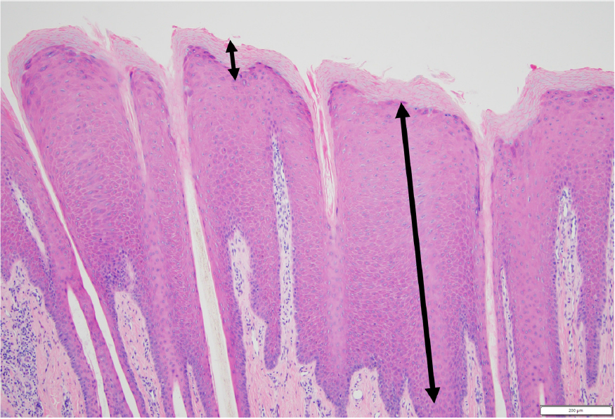
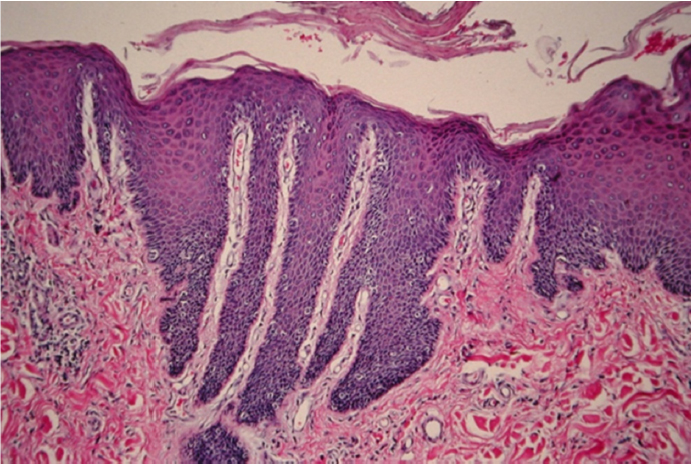
Ginn’s interpretation of the significance or cause of the lesions differs from that of Stromberg. The changes of hyperkeratosis and acanthosis were prominent in the biopsy specimens. These changes are recognized as secondary, chronic lesions and do not provide clear evidence of the primary lesion or initial injury to the skin that led to these chronic changes. The changes observed would be expected to correlate with the gross lesions of detectable irregular epidermal thickening known as lichenification. Lichenification is a term for a rough, thickened epidermis with visibly exaggerated epidermal creases or folds. Lichenified skin appears leather-like and usually is concurrent with hair loss (alopecia) (Figure 4-6). Microscopically, the stratum spinosum layer of the epidermis is thickened. There is often concurrent thickening of the stratum corneum (Figure 4-7).
___________________
3 Dr. Paul Stromberg from Ohio State University, Columbus, and Dr. Lynne Cassone of the University of Kentucky, Lexington.
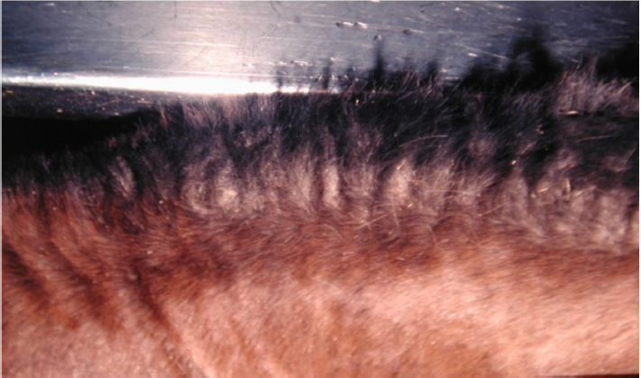
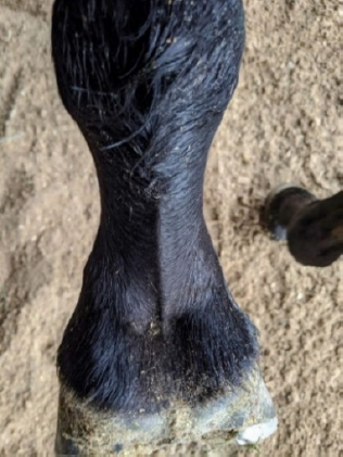
Lichenification is a pathologic change most often caused by rubbing, scratching, or some other repeated irritation of the skin. The skin changes are not incidental or insignificant and do not represent the normal character of the palmar aspect of the pastern of the horse (Figure 4-8). In addition, the subtle changes in the elastin fibers of the dermis in some horses with lichenification may be a clue to what the primary injury was. The primary injuries to the pastern of the horses in the Stromberg study or any of the TWHs presenting with lichenification of the skin of the palmar aspect of the pastern are not known (Figure 4-9). It is possible that the action devices used on the TWHs could contribute to the formation of these lesions, but this seems extreme. The caudal pastern of a horse is an area that is not very accessible to the horse, making lichenification due to some form of self-inflicted repeated injury to this area of the skin by the horse unlikely.

A long-standing federal court ruling currently limits the weight of the action devices to a maximum weight of 6 ounces, a weight limit determined not to cause injury to the pastern of the horse. The weight-limit regulation applies to action devices used during competition as well as during any training so as to eliminate any soring abuse. It is well known that action devices of weight greater than 6 ounces are used during training, so it is possible that these heavier devices could lead to the changes seen. This would still be a violation of the Horse Protection Regulations. Equine veterinarians on the committee noted that skin changes seen on the pasterns of TWHs are not observed on the pasterns of other breeds of horses (Arabians, American Saddlebreds, Morgan horses), which also train with action devices such as chains and rollers but do not wear them when shown at competitions. Action devices used in other breeds are not limited by weight and usually of lower weight than those used in TWHs. Walking horses are often trained with action devices weighing in excess of the 6-ounce action devices currently allowed for competition. The use of heavier or more cumbersome devices in training may be more likely to contribute to the formation of the lesions described in this report.
A horse included in the Stromberg study and documented as disqualified from competition at the 2015 Tennessee Walking Horse National Celebration was featured in an online article posted in 2016 that included a photograph of the horse’s pastern at the time of disqualification (Billy Go Boy Chat, 2016). The photograph of this horse shows gross lesions of erythema (redness) and swelling of the coronary band along the medial, lateral, and caudal aspect of the hoof wall. In addition, the palmar aspect of the pastern has ulceration in a V-shaped pattern in the mid region of the caudal palmar pastern of the front limb. Additional lesions of erythema and possible ulceration are present at the palmar aspect of the limb in the region of the fetlock. These acute and subacute gross lesions do not correlate with the histological findings reported in the Stromberg study which represent more chronic lesions.
Finding 4-1: Evaluation of skin samples collected from TWHs that were found to be noncompliant with the scar rule indicated variable (moderate to severe) epidermal hyperplasia (clinically evident thickening and roughness or lichenification) in the form of acanthosis (thickening of the stratum spinosum layer of the epidermis) and variable degrees of hyperkeratosis (thickening of the stratum corneum layer of the epidermis). These skin changes are not incidental or insignificant and do not represent the normal character of the palmar aspect of the horse’s pastern. In addition, skin changes seen on the pasterns of TWHs are not observed on those of other breeds of horses, which also train with action devices but usually of lower weight compared to those used on TWHs.
Finding 4-2: The changes of hyperkeratosis and acanthosis, which were prominent in the biopsy specimens, do not normally occur without a previously inflicted injury on the pasterns. These changes are recognized as secondary, chronic lesions, and they do not provide clear evidence of the initial injury to the skin leading to these changes. They are, however, expected to correlate with the grossly detectable lesions of irregular epidermal thickening known as lichenification, a pathologic change most often caused by rubbing, scratching, or some other repeated trauma to the skin.
Conclusion 4-1: The primary injury to the pastern of horses from which skin samples were collected or of any of the TWHs presenting with lichenification of the skin of the palmar aspect of the pastern is not known. It is possible that action devices alone worn by walking horses could have led to the formation of these lesions; however, this seems highly unlikely if the federal regulation limiting the weight of the action device to 6 ounces was followed.
Conclusion 4-2: More studies are needed to determine if training practices that can cause soreness in TWHs also result in lichenification. A longer-term observation of horses that are subjected to training conditions identical to TWHs training for competition but without use of any chemicals or other agents known to have been used for soring is needed. These studies might elucidate at what point, if at all, during training epidermal hyperplasia and lichenification would develop and what particular training practices would cause these conditions. It is important that observations include periodic biopsy of the palmar aspect of the pastern to check for microscopic changes.
Conclusion 4-3: Studies are also needed to determine if epidermal thickening (hyperplasia) and lichenification are solely caused by the action devices worn by TWHs. This would require observing pasterns of walking horses that were not trained for competition but were made to wear action devices under circumstances identical to TWHs in training for competition.
EVALUATION OF THE SCAR RULE CRITERIA FOR COMPLIANCE WITH THE HORSE PROTECTION ACT
The HPA and scar rule were written without any microscopic evaluation of skin lesions from horses suspected of being sore. The language of the rule was based on clinical evaluation of the skin only and has not been reviewed since its original inclusion in the Horse Protection Regulations and its implementation in 1979. Veterinary dermatopathology is now a well-recognized field of study and provides a solid framework for the accurate evaluation and characterization of skin lesions in animals. The committee believes that the rule should be revised using well-defined current medical terms that accurately describe the lesions seen today from both the clinical and histopathological standpoints.
Basis of the Scar Rule
The language of the scar rule is based on the assumptions that certain lesions exist microscopically, that those lesions can be detected by gross clinical dermatologic exam, and that the terms used in the scar rule were used appropriately. In addition, it is assumed that the rule can be interpreted and applied in a consistent manner by Animal and Plant Health Inspection Service (APHIS) veterinary medical officers (VMOs) and by designated qualified persons (DQPs) tasked with examining horses for scar rule violations. None of these assumptions hold true today, and therefore the rule as written is not enforceable. Veterinary medicine is far advanced compared with its state in 1979. In fact, the American College of Veterinary Pathologists, a specialized group of veterinarians whose focus is studying the pathologic basis of disease, was in its infancy, with its certifying examination process not available until 1978. Experts in the recognition, description, and interpretation of pathologic lesions of the skin of animals were not recognized at that time. They are today.
The first fallacy of the scar rule is the assumption that the clinical examination of gross lesions can accurately and reliably correlate with the true underlying pathologic changes in the tissue without a microscopic examination. For instance, the rule states that the fore pasterns must be free of “bilateral granulomas.” When the scar rule was written, it was common for TWHs to have very obvious skin lesions, many of which were likely a result of foreign substance injected or applied and absorbed into the skin. A granuloma is an inflammatory lesion composed of specific types of leukocytes arranged in a particular way. Granulomas most commonly form in response to foreign material and certain types of infectious agents. There is no evidence in the literature to indicate that granulomatous inflammation or granulomas have been present in the lesions of sore horses. The assumption that this type of inflammatory response may have occurred as a consequence of injection of foreign substances into the skin is reasonable, but it has never been proven to be true. In addition, this type of lesion cannot be determined to be present by the presence of clinical gross lesions alone. A microscopic evaluation of the tissue is absolutely necessary to establish the presence of granulomatous inflammation. Likewise, the use of the term “proliferating granuloma tissue” leads to confusion. In its original meaning, “proliferating granuloma tissue” may have referred to granulation tissue, but it may also have been referring to areas of granulomatous inflammation, which do not “proliferate.” Again, granulomas cannot be determined to be present by gross examination alone. The use of the word “granuloma” may have been intended to refer to the proliferation of granulation tissue which can often be recognized grossly and which corresponds microscopically to the proliferation of small capillaries and collagen-producing fibroblasts arranged in a specific pattern. Granulation tissue formation is a common finding in open wounds of horses, and it is possible to recognize granulation tissue clinically. The terms granuloma and granulation tissue are likely still used in practice to both refer to granulation tissue. Clarification of the proper use of terms in the scar rule is needed for legal
enforceability. The scar rule also refers to bilaterally uniformly thickened epithelial tissue, which is confusing. The thickening of the epidermis present in the biopsy specimens reviewed is not “uniform.” It is irregular and characteristic of lichenification.
The lesions of cardinal signs of inflammation such as edema and erythema (reddening) of tissue and also pain and the presence of moisture can likely be detected by most examiners.
The name of the rule itself, “the scar rule,” is very misleading but has been in common use for decades and refers to recognizable lesions in violation of the rule. It is still important to correct the language of the rule if it is to be enforceable in a court of law. Scars have not been documented microscopically in TWHs that have been found to be sore. A scar is an area of tissue where the normal components and organization of the tissue have been lost and replaced by fibrous connective tissue. Scars can be grossly evident, but there is no reliable documentation in the literature of a gross lesion found on a sore TWH that is compatible with a scar. Scars were very likely present in the lesions seen on sore TWHs before the enactment of the HPA. Lesions present today are more subtle, and the limited microscopic studies that have been done have not documented scars in horses determined to be in violation of the scar rule, which renders the usage of the term “scar” inappropriate.
History of Skin Lesions in Tennessee Walking Horses Suspected of Being Sore
Prior to the enactment of the HPA and the implementation of the scar rule, lesions in sore horses were grossly evident and located primarily on the anterior skin of the dorsal and palmar (caudal) pastern regions. Though not evaluated histologically, the gross lesions and history of substances applied were very suggestive of the type of injuries seen with contact irritants and most likely were characterized by the primary lesions of vesicles and secondary lesions of erosions and ulcers similar to what might have been seen with an exaggerated application of the blistering process that used to be in practice in the treatment of certain conditions of the distal limb of the horse.
After the enactment of the HPA and implementation of the scar rule, the focus on detecting a skin lesion or scar rule violation shifted to primarily involving the caudal pastern, though clearly there was still an effort to cosmetically alter the anterior surface of the pastern and coronary band region to hide more subtle evidence of injury. The reasons for the shift of focus in the location of more obvious lesions is not documented but may have occurred as weights of action devices changed and attempts were made to hide evidence of injury.
Proposal of New Scar Rule Language
A gross examination of the skin of the distal limb of the horse should still be part of the inspection process. Evaluating biopsy tissue from horses is not practical and is likely to be considered invasive, and clearly repeated biopsies could lead to tissue changes that could be confused with lesions consistent with a violation (an alternative method to study tissue changes in TWHs would be the use of ultrasonography, see Box 4-1). The language describing what constitutes a violation of the HPA should be based on what can accurately be assessed by a gross examination. Furthermore, the examination should be performed only by an experienced equine practitioner. The language of the rule should not be prescriptive and should be written so as to include any evidence of injury to the skin of distal limb. Evidence of both acute (primary) and chronic lesions should be considered evidence of an HPA violation. The committee proposes the language below as replacement for the current language in the scar rule:
A trained inspector should examine skin of the front limb of the horse from the knee (carpus) to the hoof with particular attention to skin of pastern and fetlock and the coronary band. All areas of skin from carpus to hoof of both limbs should be free of foreign substances such as dyes, hair fillers,
ointments, and other substances designed to camouflage scar rule violations during pre- and postshow inspections. Detection of previously approved substances such as lubricants during post-competition inspection does not constitute a violation. There should be no chemical smell emanating from the skin and no substance present that can be rubbed off onto the hands or a cloth. Skin should be haired with no areas of loss of hair, patchy or diffuse. There can be no swelling, redness, excoriation, erosions, ulcers, seeping of fluids, or signs of a response to chronic injury such as epidermal thickening or presence of scales. Photo documentation of lesions, identifying information about the horse, and a date should be provided for any horse determined to be or suspected of being in violation of the scar rule.
Finding 4-3: The Horse Protection Regulations and scar rule were written without any microscopic evaluation of skin lesions from horses suspected of being sore. The scar rule language was based on a clinical evaluation of the skin only and has not been reviewed since its inclusion in the regulations.
Conclusion 4-4: The scar rule language is based on the assumption that certain lesions exist microscopically and that those lesions can be detected by gross clinical dermatologic examination and also that the terms used in the scar rule were used appropriately. In addition, it is assumed that the rule can be interpreted and applied in a consistent manner by VMOs and DQPs tasked with examination of horses for scar rule violations. None of these assumptions hold true today, and therefore the rule as written is not enforceable.
Conclusion 4-5: The scar rule language needs to be based on what can accurately be assessed by a gross examination, which ideally would only be performed by an experienced equine practitioner.
RECOMMENDATION
Recommendation 4-1: Regardless of why the scar rule was written with limited information and limited expertise in pathological changes in the skin, the committee recommends that the rule be revised. The committee’s proposed language is as follows:
A trained inspector should examine skin of the front limb of the horse from the knee (carpus) to the hoof with particular attention to skin of pastern and fetlock and the coronary band. All areas of skin from carpus to hoof of both limbs should be free of foreign substances such as dyes, hair fillers, ointments, and other substances designed to camouflage scar rule violations during pre- and postshow inspections. Detection of previously approved substances such as lubricants during post-competition inspection does not constitute a violation. There should be no chemical smell emanating from the skin and no substance present that can be rubbed off onto the hands or a cloth. Skin should be haired with no areas of loss of hair, patchy or diffuse. There can be no swelling, redness, excoriation, erosions, ulcers, seeping of fluids, or signs of a response to chronic injury such as epidermal thickening or presence of scales. Photo documentation of lesions, identifying information about the horse, and a date should be provided for any horse determined to be or suspected of being in violation of the scar rule.
REFERENCES
Baird, S. 2017. Ultrasound imaging and horse health. https://www.mvsequine.com/ultrasound-imaging-and-horsehealth (accessed November 20, 2020).
Billy Go Boy Chat. 2016. Tennessee Walking Horse–“The Lickers and The Flatters,” September 13. http://billygoboy.com/2016/09/13/newly-crowned-world-grand-champion-honors-owner-keith-mcswain-slammed-by-usda-in-complaint-filed-in-washington-d-c-alleging-5-hpa-violations-aug-24-2013-july-1-2016/ (accessed July 30, 2020).
Genovese, R. L., N. W. Rantanen, M. L. Hauser, and B. S. Simpson. 1986. Diagnostic ultrasonography of equine limbs. Veterinary Clinics of North America: Equine Practice 2(1):145–226.
Hargis, A., and P. E. Ginn 2011. The integument. In J. F. Zachary and M. D. McGavin (eds.), Pathologic basis of veterinary disease, 5th ed. St. Louis, MO: Mosby-Elsevier. Pp. 972–1084.
Kettelhut, E. A., J. Traylor, and J. P. Roach. 2020. Erythema ab igne. In StatPearls [Internet]. Treasure Island, FL: StatPearls Publishing. https://www.ncbi.nlm.nih.gov/books/NBK538250 (accessed September 20, 2020).
Mlosek, R. K., and S. Malinowska. Ultrasound image of the skin, apparatus and imaging basics. Journal of Ultrasonography 13(53):212–221.
Stromberg, P. 2017. Summary report about soring in Tennessee walking horses. Unpublished manuscript, available from the Public Access Records Office of the National Academies of Sciences, Engineering, and Medicine.


