
2
Tissue Homeostasis, Inflammation, and Repair
A keynote presentation on tissue homeostasis, inflammation, and repair was delivered by Ruslan Medzhitov, Sterling Professor of Immunobiology at Yale University School of Medicine and an investigator at Howard Hughes Medical Institute, at the beginning of the workshop. Tissue homeostasis involves the minimum cell components that constitute a tissue, a
feedback circuit within the tissue, and the ways that cells within the circuit respond to environmental pressures such as available space, growth factors, cytokines, oxygen, and tension. Medzhitov reviewed the established fundamentals about these processes and highlighted gaps in our current understanding as a way to set the stage for subsequent workshop sessions on tissue regeneration.
TISSUE HOMEOSTASIS
Much is known about cellular and organismal homeostasis, but little is understood about tissue homeostasis,1 said Medzhitov. Cellular homeostasis involves what some biologists refer to as “cell stress responses,” where cellular sensors detect levels of oxygen, glucose, protein, and other variables; when a variable changes, it initiates an appropriate adaptive response. At the systemic level, for example, oxygen stress responses to hypoxia are regulated by erythropoietin and red blood cell production, while vascularization regulates the response at the tissue level. Although angiogenesis and hypoxic response are relatively well understood, he said, most variables of tissue homeostasis are not. A number of microenvironmental variables are involved in maintaining tissue homeostasis (see Table 2-1). These include (1) oxygen and nutrients; (2) cell number and composition; (3) the composition and mechanical properties of extracellular matrix (ECM); and (4) interstitial fluid volume, pH, osmolarity, and metabolic waste products. The mechanisms that maintain these variables within tissues are unknown, with a few exceptions such as hypoxia. Medzhitov remarked that it is not the complexity of the mechanisms that impedes understanding of tissue homeostasis, but rather a lack of research in these areas.
COMMON FEATURES OF TISSUE ORGANIZATION
Although tissue types from different organs have superficial differences in appearance, biologists have traditionally understood that all tissue types share common themes in tissue architecture and design principles. This idea is based on the understanding that a biological problem is solved by evolutionary processes, and that solution is maintained to address related problems, Medzhitov explained. For example, different tissues face similar problems, such as determining cellular composition in terms of the cell types present, their amounts and proportions, and their spatial distribution. Some cells must also be able to expand on demand, such as immune cells during an inflammatory response. He posited that early in the evolution
___________________
1 Tissue homeostasis is a “collection of circuits that regulate specific variables within the tissue environment” (Meizlish et al., 2021).
of animals, a solution was developed, and in turn, all human tissues are variations on a theme related to that solution. Yet, that theme is not known. This knowledge gap limits the ability to develop rational approaches to targeting diseases at the tissue level; it is important to appreciate how little is understood at this biological level, he emphasized.
| Regulated Microenvironmental Variables | Manipulated Processes |
|---|---|
| Oxygen and nutrients | Local blood profusion level; angiogenesis |
| Cell number and composition | Cell proliferation; cell death; cell migration |
| ECM level, composition, and stiffness | Production and degradation of ECM components; collagen crosslinking |
| Interstitial fluids volume, pH, and osmolarity; metabolic waste products | Vascular permeability; lymphatic drainage; perfusion level; (lymph) angiogenesis; acid–base control; solute transport |
NOTE: ECM = extracellular matrix.
SOURCE: Medzhitov presentation, November 2, 2021.
TISSUE ORGANIZATION AND COMPOSITION
Basic tissue organization includes multiple cell types and relies upon the extracellular matrix. Tissue organization raises many questions, such as how cells “know” to exist in specific locations and what relationships there are among different cell types. Medzhitov noted that some cell types are more important than others. For example, if lymphocytes were removed from tissues, some functionalities would be lost but the overall tissue architecture would be preserved. In contrast, tissue structure is lost with the removal of fibroblasts. Thus, some cell types are more foundational than others. The question that then arises is how to define the minimal composition of tissue necessary for normal architecture, said Medzhitov. Evolutionary history can inform the answer because the simplest animals like placozoans or sponges have at least two types of cells: epithelial cells and mesenchymal cells. Together, epithelial-mesenchymal modules constitute the primordial units, or building blocks, of tissue organization.
Epithelial and mesenchymal cell types have specific relationships associated with the flow of information between them, Medzhitov explained. For example, stromal mesenchymal cells contain positional information about their location along the body’s axis. They produce morphogen signals that act on epithelial cells to determine their cell fate (i.e., the different types of
epithelial cells they become). This relationship between mesenchymal and epithelial cells generalizes to other cell types—that is, niche cells control the cell fate of stem cells or, in the immune system, dendritic cells control that of T cells. Furthermore, the relationship hints at the fundamental rules of tissue organization, he said. Some cells are more similar to mesenchymal cells in that they contain information, and some cells are more like epithelial or functional cell types in that they have various fate choices.
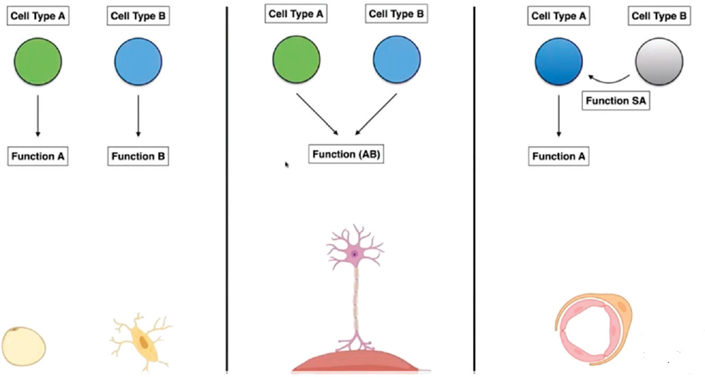
CELLULAR DIVISION OF LABOR
Cell types also have relationships that enable them to perform necessary functions, Medzhitov noted. Cellular division of labor can be conceptualized in different ways (see Figure 2-1). Classically, the division of labor consists of two cell types that each specialize in a different function. For instance, adipocytes and osteocytes perform different functions independently of one another. Another functional relationship occurs when two cell types contribute to the same function, and both types are required to perform the function. For example, motor neurons and skeletal muscle cells are both needed to execute the function of contractility. These cell combinations are referred to as functional units.
A third form of cellular division of labor occurs when one cell type is specialized to perform a function and another cell type specializes to support the function of the first cell. For instance, a neuron is a functional cell supported by a glial Schwann cell. A Schwann cell has no functional meaning without a neuron; its meaning is predicated upon supporting a neuron. In contrast, neurons can operate without Schwann cells, and many of them do. Medzhitov added that, by analogy to client and chaperone proteins, functional and supportive cells can also be referred to as client cells and accessory cells. Applying this understanding to tissue composition underscores that some cells are more foundational than others. Some cells are functional client cells responsible for the primary function of the tissue while other cells perform supportive functions. Finally, some cells provide information relevant to fate decisions by other cells through directional signaling to those cells.
MINIMAL TISSUE UNITS AND GROWTH FACTOR PRODUCTION
The minimal composition of tissues in vertebrates comprises four cell types that form “minimal tissue units,” Medzhitov said. The first is responsible for the core function of the tissue and is the most common cell type of a tissue. Examples include tissue-specific cells like epithelial cells in the lung, hepatocytes in the liver, and neurons in the brain. Three other
SOURCE: Medzhitov presentation, November 2, 2021.
cell types perform supportive functions: microvascular endothelial cells, fibroblast-like stromal cells, and tissue-resident macrophages. These three cell types are nearly universal in all tissues and perform supportive functions, such as delivering oxygen and nutrients, producing ECM, providing defense, and maintaining homeostasis. The primary design of a tissue consists of these four cell types, but there are exceptions; for example cartilage, he noted. Although not responsible for a primary function of the tissue, the supportive cell types are necessary for the primary cell type to perform its function optimally. The supportive cell types follow their genetically encoded instructions to support the client cell’s functionality even if the client cell becomes cancerous. Therefore, the three supportive cell types are universally present in solid tumors, dutifully performing their functions despite inadvertently promoting tumor growth, Medzhitov described. This feature of supportive cells is important to consider in determining which cell categories to target for regeneration, he added.
Every normal, nontransformed cell type requires specific growth factors to survive and proliferate, and the number of cells in a given location is determined by the local availability of growth factors. Thus, the cellular composition of tissues is dictated by the local availability of lineage-specific growth factors. The question of tissue design, Medzhitov said, can be translated into a question of what controls the local provision of growth factors. For example, macrophages will not be found in tissue devoid of colony stimulating factor-1 (CSF-1), and fibroblasts make CSF-1. Similarly, macrophages and endothelial cells make platelet-derived growth factor (PDGF) to support stromal fibroblasts. The presence of cell types in the correct quantity and location depends on appropriate production of corresponding growth factors, he explained. Cells have rules and mechanisms to ensure the production of growth factors in the right amounts and locations, but these rules and mechanisms are largely unknown. Knowledge of the rules that regulate growth factor production could be applied to support the purposes of homeostasis, regeneration, or cell therapy, he suggested.
Macrophage-Fibroblast Two-Cell Circuit
Typically, the growth factor for one cell type is produced by another, said Medzhitov. That is, cell X makes growth factor for cell Y, and the amount of growth factor determines the quantity and location of cell Y within the tissue. Generally, it is not yet understood how cells determine how much growth factor to make to ensure appropriate cell composition for the tissue. In studying macrophages and fibroblasts, Medzhitov and his colleagues found that the cell types exchange growth factors according to specific principles (Adler et al., 2018; Zhou et al., 2018). Fibroblasts produce CSF-1 for macrophages, and macrophages make PDGF for fibroblasts.
A particular regulatory circuit establishes stable provision of growth factors between macrophages and fibroblasts (see Figure 2-2). Space availability limits growth of fibroblasts while growth factor availability limits macrophages. Furthermore, this circuit design is stable to perturbations, and the concept of a minimum stable circuit is likely generalizable to other systems and cell types, including immune cells, Medzhitov explained (Zhou et al., 2018). He added that the source of growth factor for the client cell type could be regulated by a principle such as extrinsic space restriction.
TISSUE OR ORGAN SIZE CONTROL
Medzhitov outlined two coexisting paradigms for regulating compartment (e.g., tissue or organ) size. The first paradigm centers on space availability. In this model, cells in the tissue compartment proliferate until they fill all available space, at which point proliferation ceases. The in vitro counterpart is contact inhibition. This scenario applies to some cells—such as fibroblasts and endothelial cells—but not to others, including hematopoietic cells, lymphocytes, and granulocytes. The second paradigm centers on growth factor availability and proposes that tissues locally produce appropriate amounts of growth factor. An abundance of growth factor induces cell proliferation, and if cells proliferate beyond a level sustainable by the amount of growth factor, excess cells die to reach the appropriate compartment size.
Given that fibroblasts are limited by space availability and macrophages are limited by growth factor availability, these two regulatory paradigms correspond exactly with the two cell types. Moreover, Medzhitov added,
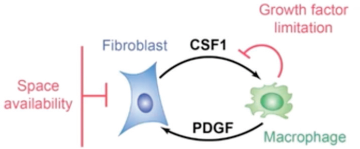
NOTE: CSF1 = colony stimulating factor 1; PDGF = platelet-derived growth factor.
SOURCES: Medzhitov presentation, November 2, 2021; adapted from Zhou et al., 2018.
he and his colleagues found that the two paradigms are mechanistically coupled, in that space availability for fibroblasts thereby controlled growth factor production for macrophages. Through a homeostatic or density-sensitive enhancer, fibroblasts sense available space and produce the requisite amount of CSF-1 growth factor to generate the number of macrophages that correspond to the available space detected by fibroblasts. In this way, the two paradigms are coupled, he explained.
Macrophage growth factor CSF-1 also has a ubiquitous inflammation-sensitive enhancer that is controlled by nuclear factor kappa B, or NF-kB. This inflammatory enhancer is independent of cellular density and enables macrophages to expand as necessary to support the inflammatory response. This principle of growth factor production—in which one cell type senses the tissue microenvironment and produces growth factor to regulate another cell type—can be further generalized, Medzhitov stated. One well-known example is that oxygen sensed through the hypoxia-inducible factor-1 pathway regulates vascular endothelial growth factor, or VEGF, which in turn controls the appropriate numbers of endothelial cells. The general rule seems to be that one cell type, which senses the microenvironment, is linked to population size control of another cell type, Medzhitov said.
TISSUE REPAIR AND REGENERATION
Tissues can be described as labile, stable, or permanent based on the type and degree of cell turnover, Medzhitov explained. Labile tissues have stem cells like epithelial cells and hematopoietic cells, and differentiated cells in the tissue are continuously renewed by stem cells. Although labile tissues are easily damaged, they are also easy to repair by way of regeneration, in which lost cells are simply replaced, he said. Stable tissues have some capacity for regeneration if the damage is limited, but if the damage is extensive, repair occurs by fibrosis. In stable tissues, stem-cell–based renewal is not typical; rather, regeneration happens via mitosis of terminally differentiated cells, such as hepatocytes, fibroblasts, and pancreatic beta cells. Comprised of cells like neurons and cardiomyocytes, permanent tissues can only repair by fibrosis, making these tissues vulnerable to degenerative disease. These three classes of tissues have different rules that govern how supportive cells assist functional client cells, how cell population sizes are controlled, and how the tissues repair. He added that the optimal regenerative approach will, therefore, depend on the category to which the target cell belongs.


