
The objectives of the third session of the workshop were to (1) examine what “proper healing” looks like at the level of the local environment and discuss relevant research gaps and (2) consider the effects of aging, gender, and other variables and pathological changes on the local environment, endogenous repair, and wound healing. Steven Becker, program director of the Structural Biology and Molecular Applications Branch at the National Cancer Institute at the time of the workshop, moderated the session.
REVERSING AGING: PRO-INFLAMMATORY METABOLITE PROSTAGLANDIN E2 AUGMENTS MUSCLE REGENERATION
Helen Blau, Donald E. and Delia B. Baxter Foundation Professor and director of the Baxter Laboratory for Stem Cell Biology at Stanford University, discussed the discovery that prostaglandin E2 (PGE2), an inflammatory metabolite, is crucial to muscle regeneration. PGE2 also plays a major role in aging, and targeting its degradation can both ameliorate aging and promote regeneration, effectively reversing the aging process in muscle, she said. Regenerative medicine questions whether aging must entail debility that hampers quality of life in old age, Blau observed. Life expectancy has increased over the past 15 years for both men and women (Bellantuono, 2018). However, it is life span—rather than healthspan—that is increasing, so people spend more years living with chronic disease. Men born in 2014 are expected to experience three more years with chronic disease than are those born in 2006. Blau highlighted the goal of improving quality of life and healthspan through regenerative medicine.
Combating Muscle Wasting
Muscle supports a high quality of life because it is central to all life activities, from dancing in a ballet to using diaphragm muscles while sitting, said Blau. Aging can involve a devastating loss of muscle mass and strength, where the average human loses 10 percent of their muscle mass per decade after the age of 50 (Hunt et al., 2015). One third of the population aged 80 years or older is affected by the debilitating loss of muscle mass and strength known as sarcopenia (Looker and Wang, 2015). Loss of muscle mass has severe consequences (Burns et al., 2016). As people become frail, they are less able to perform everyday tasks, including walking and rising from a chair. Falls become more common, resulting in $31 billion in annual injury treatment costs for nonfatal falls (Burns et al., 2016). As tasks of daily living become more difficult, people become more dependent and may require institutionalization, which carries further high health care costs. Sarcopenia is also associated with increased mortality, Blau noted.
Muscle stem cells (MuSCs) are essential to muscle regeneration, and they give rise to two key methods to combat muscle wasting, including (1) MuSC stimulation to enhance regeneration and (2) muscle myofiber rejuvenation, Blau explained. Both approaches are mediated by the metabolite PGE2. MuSCs are resident in muscle tissue and are dedicated to the repair of muscle throughout life. Located in niches along the basal lamina of the myofiber, MuSCs are in a quiescent state until injury occurs. When the muscle is injured, the stem cells are activated and express myoblast determination protein 1, also known as MyoD. The MuSCs then become committed progenitors that express fusion proteins that mediate fusion with the damaged myofibers, replenishing them with new nuclei and, consequently, new DNA. Interestingly, this is the same population of stem cells which Alexander Mauro correctly postulated that he saw on electron micrographs and which he referred to as satellite cells in 1961, Blau noted (Mauro, 1961).
Effect of Prostaglandin E2 on Muscle Repair
A natural inflammatory regulator, PGE2 is one of the body’s first responses to injury. Injury triggers waves of sequential cellular activity, beginning with inflammation, followed by fibroadipocytes and fibroblasts and then by activation of MuSCs. Blau and her team reasoned that a regulator in this cascade must strongly influence the function of MuSCs and that aging alters this effect (Blau et al., 2015). Andrew Ho and Adelaida Palla, scientists from the Baxter Laboratory for Stem Cell Biology at Stanford University, conducted a bioinformatic analysis of the transcriptome of activated stem cells acutely post-injury (Ho et al., 2017). They culled a common gene list of activated MuSC transcripts and used Ingenuity Pathway Analysis to focus on transmembrane and receptor genes (Ho et al., 2017). Ho and Palla identified EP4, an E-type prostanoid receptor for PGE2, as one of the most common receptors in the category of inflammation, suggesting an important role for PGE2 in stimulating MuSC function and repair. Despite its important role, PGE2 may be understudied because it is a metabolite and would, therefore, not appear on a transcriptome, Blau explained. PGE2 is derived from arachidonic acid via several enzymatic steps. Although PGE2 can work on any one of four EP receptors, EP4 is by far the most prevalent in MuSCs and muscle fibers, and EP4 signals through cyclic adenosine monophosphate (cAMP) (Ho et al., 2017). In addition, nonsteroidal anti-inflammatory drugs (NSAIDs) such as indomethacin and ibuprofen block cyclooxygenase enzymes (COX-1 and -2), which in turn block the endogenous synthesis of PGE2 after injury, Blau noted (Ho et al., 2017).
Loss of Prostaglandin E2 Signaling in Muscle Stem Cells Impairs Muscle Regeneration
To determine whether this pathway is crucial to the function of MuSCs, Blau and her colleagues conducted a conditional genetic ablation of the receptor EP4 on MuSCs in a mouse model using the promoter of paired box 7 (PAX7), a transcription factor that is a hallmark of the stem cell and drives floxing of EP4 in the knockout model. The researchers performed an injury by injecting notexin, a snake venom toxin commonly used in the field to cause muscle injury. Several weeks later, strength was measured using a foot pedal measurement of displacement. They found that when the stem cells could not sense PGE2 due to a lack of EP4 receptors, the mice experienced a 50 percent decrease in strength, both in twitch force and tetanic force (Ho et al., 2017). Therefore, this pathway is extremely important for enabling stem cells to proliferate, expand, and repair the damage, and young mice became weaker after injury due to their inability to repair muscle damage, Blau explained.
NSAID Treatment Decreases Endogenous Prostaglandin E2 Signaling after Injury and Impairs Muscle Strength
Blau and her team further examined the role of PGE2 signaling through loss-of-function experiments. For instance, using another mouse model with PAX7-driven luciferase expression, they performed a muscle injury and administered an NSAID, the equivalent of an ibuprofen injection (Ho et al., 2017). Reduced bioluminescence, a readout of luciferase, correlates with fewer stem cells due to decreased proliferation and expansion in comparison to mice that did not receive the NSAID, Blau described. Twitch force also saw a remarkable decline, she noted. The New York Times highlighted this study in an article entitled “Bring on the Exercise, Hold the Painkillers” (Reynolds, 2017). Although many people take ibuprofen after working out to decrease achiness, taking an NSAID negates the benefit of exercise, and the injury associated with exercise is part of building muscle and muscle strength, Blau remarked. The pathway is essential to proper stem cell function, and if PGE2 signaling is blocked, the cells lose much of their ability to divide, repair injury, and build strength, she said.
Globally Increasing Muscle Function with Prostaglandin E2
Their findings about the local effects of PGE2 led Blau and her colleagues to wonder whether global PGE2 levels could be increased and what effect that would have on muscle function. While working at the Baxter Laboratory for Stem Cell Biology at Stanford University,
Adelaida Palla, Meenakshi Ravichandran, and Yu Xin Wang discovered that 15-hydroxyprostaglandin dehydrogenase (15-PGDH), the degrading enzyme of PGE2, is a novel molecular determinant of muscle aging and a regulator of prostaglandins that is crucial to muscle function (Palla et al., 2021). Furthermore, they found that 15-PGDH is a hallmark of aging and can be targeted for rejuvenation. Given the muscle weakness associated with aging, Blau and her team reasoned that PGE2 might decline with age. By measuring levels of PGE2 and prostaglandin D2 (PGD2) by mass spectrometry, they found that these metabolites decreased by approximately 30 percent with aging (Palla et al., 2021). They hypothesized that this decline might result from an elevation in the enzyme that degrades these prostaglandins. Indeed, a comparison of 15-PGDH levels in young and aged mouse tissues revealed that the enzyme not only increased in aged muscle, but also in the colon, spleen, skin, and heart (Palla et al., 2021). Thus, 15-PGDH appears to be a hallmark of aging in multiple tissues. The researchers also mined a publicly available microarray database for 15-PGDH expression to determine whether their finding held for human tissue, and they discovered higher levels of the enzyme in aged humans as well (Palla et al., 2021). Blau noted that 15-PGDH expression—like all hallmarks of aging—is on a spectrum, with some people being biologically younger than their peers.
To test whether targeting the degradation enzyme, 15-PGDH, would increase PGE2 levels, researchers used a small molecule inhibitor, SW033291, and administered daily intraperitoneal injections of the inhibitor to aged mice for one month, Blau explained. This resulted in the degrading enzyme, 15-PGDH, decreasing by approximately 30 percent and a consequent twofold increase in PGE2 and PGD2, constituting a return to youthful levels (Palla et al., 2021). The inhibition of 15-PGDH and the resulting increase of PGE2 were associated with a significant increase in muscle mass in the gastrocnemius, tibialis anterior, and soleus muscles, and therefore had an effect on both fast- and slow-contracting muscle types (Palla et al., 2021). Furthermore, muscle strength and force relative to baseline also increased in both young and aged mice. Moreover, endurance, measured by time to exhaustion on a treadmill, also significantly increased, which presumably involves more than simply skeletal muscle, remarked Blau. In order to ensure that these results were specific to 15-PGDH, the experiment was replicated using a genetic knockdown of 15-PGDH with a short hairpin RNA, and the same results were found with increases in muscle mass, muscle strength, and force relative to baseline (Palla et al., 2021). Utilizing CO-Detection by indEXing (CODEX) imaging, Blau’s team found that myofibers and macrophages were the source of 15-PGDH in aging. Accordingly, the researchers overexpressed this enzyme in young mouse models, and muscle mass, strength, and force declined within a month. Remarkably, muscles shrank and weakened in response to overexpression of a single enzyme, Blau emphasized. When
15-PGDH increased, PGE2 and PGD2 decreased, further validating earlier findings. These experiments demonstrate that 15-PGDH is a pivotal regulator of aging and rejuvenation, she said.
15-PGDH Inhibition Mechanism of Action
To determine the mechanism responsible for 15-PGDH inhibition, Blau and colleagues compared heatmaps of gene expression for three vehicle-treated mice and three mice treated with a small molecule inhibitor (SW033291) for one month (Palla et al., 2021). The mice injected with SW033291 showed a marked down-regulation of ubiquitin ligases, which are enzymes responsible for protein degradation in aging muscle. Researchers also found a decline in transforming growth factor beta (TGF-beta) signaling, including myostatin, which is a common target of therapeutics pursued in biotechnology and pharmaceutical companies for attempting to increase muscle mass (Palla et al., 2021). The results suggest that PGE2 is upstream of myostatin and that inhibition of the enzyme 15-PGDH inhibits deleterious pathways responsible for muscle degradation, Blau explained. These findings were also validated by polymerase chain reaction (PCR) testing, she noted. In addition, researchers found that inhibiting 15-PGDH spurred an increase in mitochondrial function. Heatmaps of gene expression indicated dramatic upregulation of mitochondria-related gene expression, the effects of which can be observed on an electron micrograph. Aged mouse muscle tissue featured distended, vacuous, dysfunctional mitochondria. After aged mice received one month of treatment, muscle tissue was remodeled to a youthful structure with intact myofibrils and condensed mitochondria in orderly pairs, described Blau. Quantitative analysis revealed that the autophagy protein p62 increased with treatment and is likely largely responsible for promoting mitochondrial biogenesis and restoring mitochondrial function. Additionally, an examination of Krebs cycle components indicated that citrate synthase, succinate dehydrogenase, and membrane potential all decline with aging and are restored with 15-PGDH inhibition treatment. Therefore, inhibiting the degrading enzyme restored mitochondrial function, increased mitochondrial numbers, and improved function in electron transfer and energy production, she added.
Blau concluded by emphasizing that (1) prostaglandin signaling is critical for muscle maintenance, regeneration, and rejuvenation and (2) a small molecule inhibitor of 15-PGDH has pleiotropic beneficial effects through enhancing PGE2. She provided an overview of key findings on the role of PGE2 on muscle fiber and MuSC function (see Box 5-1). PGE2 is an essential inflammatory metabolite that is required for MuSC expansion and engraftment. Inhibiting 15-PGDH enhances PGE2, rejuvenating myofibers and stem cells. Elevated 15-PGDH, a key degrading enzyme, is a novel
hallmark of aging, and it can be targeted in a physiologic way. Modulating the levels of 15-PGDH twofold, to levels present in muscles of young mice, has a beneficial effect and holds therapeutic potential for countering sarcopenia and other conditions.
BIOMATERIALS FOR MODELING IMMUNE MEDIATION IN WOUND HEALING
Erika Moore, Rhines Rising Star Larry Hench Assistant Professor at the University of Florida Department of Materials Science and Engineering, discussed the use of biomaterials for modeling immune cell mediation in wound healing. The inherent variability observed in wound healing creates an opportunity to use biomaterials to mimic differences in wound-healing environments. Minor injuries are typically met with pro-healing wound repair in which a small scar may be present but most tissue function is recovered, she explained. Conversely, dysregulated wound healing can result in keloid or hypertrophic scars. Pro-fibrotic wounds feature thick scar tissue and demonstrate the differentials that persist in wound healing. A patient-centered perspective would facilitate a better understanding of variability in wound healing because patients have different ancestral backgrounds, age contributors, experiences of trauma, and physical activity that can all contribute to the outcome in wound healing, Moore suggested.
B-Cell Involvement in Wound Healing
To investigate variability in wound healing, Moore and her colleagues assessed immune cell coordination and contribution to the injury response. Multiple cell types orchestrate wound healing via cell-to-cell communications. B cells are known for their role in responding to vaccines and creating antibodies, enabling the immune system to recognize the antigen or stimulant in future exposures and develop an immune response against it; however, studying the role of B cells in wound healing is relatively controversial, Moore remarked. Yet, a previous study demonstrated that the presence or absence of B cells yielded differences in wound healing both early in the process and at subsequent points in the healing timeline (Iwata et al., 2009). In dermal skin wound injuries, B cells orchestrated healing in combination with other immune cells present at the site, resulting in more complete healing than in injuries in which B cells were absent (Iwata et al., 2009). These findings indicate that the role of B cells extends beyond antibody presentation and secretion to include critical involvement in wound healing, noted Moore. Furthermore, the dysregulation or dysfunction of B cells is associated with many diseases, including autoimmune disorders. She and her team sought to profile the connection between the immune response at the local tissue site and the systemic or whole-organism response and to learn how B cells operate during wound healing.
Biomaterials to Manipulate Wound Healing
Moore and her team used biomaterials to create model systems of prohealing and pro-fibrotic wounds to learn how B cells respond differently in these wound environments. In an effort to control the B-cell responses, the researchers leveraged biomaterial designs in mimicking wound-healing environments, utilizing pro-healing materials that promote wound healing or pro-fibrotic materials that encourage fibrosis or scar formation. Through these two types of biomaterials, the wound-healing process could be manipulated in order to assess the B-cell response in each of these environments, explained Moore. Researchers induced muscle injury in mice, implanted biomaterial directly into the wound, and interrogated the injury sites. In addition to interrogating the local response, they also examined both regional responses that included the lymph node and systemic responses that included the peritoneum and spleen. Moore emphasized that they sought to understand how the local environment and presence of specific biomaterials skew response toward healing or fibrosis and how that informs the entire systemic B-cell response.
The pro-healing materials promoted early recruitment of immature B cells to the wound sites, Moore said. In the control group, the average
B-cell count was below 5,000 on post-injury day 3 and slightly above 5,000 on day 5. In contrast, the average B-cell count in mice implanted with pro-healing material was above 10,000 on day 3 and increased to more than 15,000 on day 5 (a threefold increase over the injury control group) (Moore et al., 2021). Furthermore, the pro-healing materials promoted a regional response, with B cells generated at the local lymph nodes of the mice after injury. At one week post-injury, germinal centers had formed in the lymph nodes, indicating maturation of B cells in the mice implanted with pro-healing biomaterial. The generation of germinal centers did not appear in the control group or in the mice implanted with pro-fibrotic material, emphasized Moore. Therefore, the pro-healing material induced specific recognition and antigen response in B cells, beyond what occurs for injury alone.
Conversely, pro-fibrotic biomaterials were found to recruit mature B cells at later stages of wound healing, said Moore. Researchers studied the effects of mature B cells by conducting a B-cell knockout in mice and implanting pro-fibrotic biomaterial. Measurements of collagen and alpha-smooth muscle actin levels in mice with B cells indicated relatively high expression of these fibrosis-related markers. When scientists removed the mature B cells, the expression of both collagen and alpha-smooth muscle actin decreased (Moore et al., 2021). Thus, removing mature B cells reduced fibrosis and scar formation in the pro-fibrotic biomaterial environment, she stated.
B Cells in Wound Healing
All people exist on a spectrum of healing, observed Moore. The ability to heal well is influenced by the tissue in which injury occurs, background, age, and other factors. This research is the first of its kind to test B-cell function in wound healing with the use of biomaterials, she remarked. Pro-healing materials were found to induce early recruitment of immature B cells, promoting healing. Pro-fibrotic materials recruited mature B cells, resulting in fibrosis. In turn, a reduction in fibrosis occurred with the removal of mature B cells from the injury site. Moore highlighted a dichotomy in the roles that B cells can play depending on their antigen-mature state, the maturity of the B cells, and on the timing of their recruitment to the injury site.
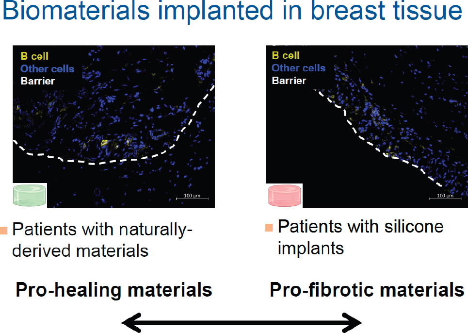
Moore and her colleagues investigate wound healing with the goal of achieving clinical relevance. One of those efforts is to study how B-cell response might alter or augment the body’s acceptance of biomaterials implanted into breast tissue. Comparing human biopsies of patients implanted with naturally derived, pro-healing material with those of patients with silicone implants has revealed that the B-cell response differs
in orientation and spread pattern between these two patient groups (see Figure 5-1). Understanding and classifying how biomaterials used in the clinic inform the immune response, and particularly the B-cell response, provides insight on the ability to regenerate, she remarked.
Research Gaps in Understanding Wound Healing
Gaps in understanding persist in this area, Moore noted. For instance, it is unclear how B cells adapt in the wound environment. Additionally, researchers do not know whether the B-cell role either in local tissue response or in secreting antibodies informs the systemic response. Moore remarked on her interest in understanding how to connect the local injury response to systemic immunity by linking the B-cell responses at the local and systemic response sites. Furthermore, research that leverages biomaterial models could elucidate patient-specific wound healing, she added. If biomaterials can mimic different wound-healing conditions and microenvironments of vulnerable populations, the contribution of patient variability—including ancestry, age, biological sex, and history of trauma—in wound healing could be interrogated.
SOURCE: Moore presentation, November 2, 2021.
ENDOGENOUS PRO-RESOLUTION AND PRO-REGENERATIVE MECHANISMS IN PERIODONTAL TISSUE
George Hajishengallis, Thomas W. Evans Centennial Professor in the Department of Basic and Translational Sciences at the University of Pennsylvania, characterized periodontitis as a chronic inflammatory disease that leads to the destruction of tissues that surround and support the teeth—the gingiva, periodontal ligament, and alveolar bone. As periodontitis progresses, loss of attachment to the teeth occurs and bone becomes severely diminished, which can lead to tooth loss. Severe periodontitis is the sixth-most-prevalent inflammatory condition worldwide, affecting 10 percent of the adult population (Kassebaum et al., 2014). Furthermore, periodontitis is associated with increased risk of systemic comorbidities, including cardiovascular disease, Alzheimer’s disease, and diabetes (Hajishengallis and Chavakis, 2021). Periodontitis also poses a significant economic burden, costing the United States approximately $154 billion in annual direct and indirect costs (Botelho et al., 2021). Hajishengallis and his team work toward the goal of promoting both resolution of inflammation and tissue regeneration in the periodontal tissue. To this end, they use a combination of in vitro mechanistic models, animal models, and human studies, including a recent phase IIa clinical trial that showed that a complement C3 inhibitor can promote resolution of periodontal inflammation in human patients (Hasturk et al., 2021).
The Functions of Developmental Endothelial Locus-1
Developmental endothelial locus-1 (DEL-1) is a molecule believed to stimulate endogenous resolution and regenerative pathways, said Hajishengallis. A secreted 52-kilodalton protein, DEL-1 consists of three epidermal growth factor (EGF)-like repeats in the N terminal and two discoidin I–like domains in the C terminal of the molecule (Yuh et al., 2020). The discoidin I–like domains interact with phospholipids, whereas the EGF-like repeats are responsible for interactions with various integrins, transmembrane receptors that bind to the extracellular matrix (Kourtzelis et al., 2019). DEL-1 is expressed primarily by tissue-resident cells, including endothelial cells, neuronal cells, and mesenchymal stem cells (MSC) in the periodontal ligament and bone marrow, and by certain macrophage subsets. Unfortunately, the expression of DEL-1 decreases substantially with aging in both humans and in mice, he noted. DEL-1 was identified as an important molecule with the discovery that it can bind lymphocyte function–associated antigen 1 (LFA-1) on leukocytes such as neutrophils, explained Hajishengallis. DEL-1 blocks the interaction of LFA-1 with the endothelial intercellular adhesion molecule 1, known as ICAM-1 (Choi et al., 2008).
This interaction is important for circulating neutrophils to adhere onto the endothelium and is a prerequisite step for their transmigration, he added.
When DEL-1 is secreted by endothelial cells, it can regulate the recruitment of inflammatory cells into underlying tissues. Hajishengallis and colleagues showed that DEL-1 could confer protection against periodontitis in both mice and nonhuman primates (Eskan et al., 2012; Shin et al., 2015). They extended these observations beyond periodontitis to other disease models by showing that DEL-1 can protect against multiple sclerosis by inhibiting inflammation (Choi et al., 2015). Recently, they demonstrated that DEL-1 is also protective against rheumatoid arthritis (Wang et al., 2021). He emphasized that location influences the functional role of DEL-1. When DEL-1 is expressed by endothelial cells, its job is to regulate neutrophil recruitment. However, when DEL-1 is produced by macrophages, the protein promotes efferocytosis and inflammation resolution because it can serve as a molecular bridge to facilitate the interaction of apoptotic neutrophils with macrophages (Kourtzelis et al., 2019). Specifically, DEL-1 binds phosphatidylserine (PS) on apoptotic cells through its discoidin I–like domains and beta 3 integrin on macrophages through an arginine-glycine-aspartic acid (RGD) motif of the second EGF repeat, thereby promoting the ability of macrophages to engulf apoptotic cells (Kourtzelis et al., 2019). Hajishengallis explained that DEL-1–mediated uptake of apoptotic neutrophils has a function beyond waste disposal. That is, the efferocytic macrophage becomes pro-resolving and begins secreting transforming growth factor (TGF) beta, resolvins, and other factors important for resolving inflammation. This is the mechanism by which DEL-1 promotes inflammation resolution in periodontitis (Kourtzelis et al., 2019).
Role of Developmental Endothelial Locus-1 in Periodontitis
Researchers have found that DEL-1 can also induce regeneration of bone in periodontitis through an independent mechanism (Yuh et al., 2020). Hajishengallis and his colleagues have used a mouse model in which they induced experimental periodontitis by ligation at the molar teeth, causing accumulation of the biofilm that produces inflammation and subsequently results in alveolar bone loss. At day 10 the ligature was removed, facilitating transition to the resolution phase. Wild-type mice regenerated bone during the resolution phase, whereas the DEL-1–deficient mice did not (Yuh et al., 2020). When DEL-1 knockout mice received a local administration of recombinant DEL-1, the ability to regenerate bone was restored. Surprisingly, the effect of bone regeneration was restored even with the local administration of a segment of DEL-1 consisting of only EGF-like repeats, suggesting that the mechanism depends on EGF-like repeats and does not require the discoidin I–like domains, Hajishengallis said.
A key implication of these data is that the mechanism by which DEL-1 regenerates bone is independent of its ability to promote efferocytosis and resolution, stated Hajishengallis. Inflammation resolution is also important for bone regeneration, but it is significant that DEL-1 carries out these two functions independently, he emphasized. The entire DEL-1 molecule is required for efferocytosis, with one part binding to the macrophages and the other part binding to the apoptotic neutrophils (Kourtzelis et al., 2019). However, the EGF-like repeats alone were sufficient for bone regeneration (Yuh et al., 2020). Hajishengallis and his team also found that the RGD motif in the second EGF-like repeat is critical for bone regeneration. Using a transgenic mouse model with a mutation in the RGD motif of DEL-1 that substituted glutamic acid for aspartic acid, they demonstrated that the mice were unable to regenerate bone during the resolution of periodontitis without this vital domain (Yuh et al., 2020).
In vitro mechanistic studies were used to confirm the in vivo findings, said Hajishengallis. Experiments using a culture model with calvarial osteoblastic cells confirmed that the RGD motif and EGF repeats mediate the ability of DEL-1 to induce osteogenic differentiation and calcified nodule formation (Yuh et al., 2020). Researchers used MC3T3-E1 osteoblastic progenitor cells to dissect the pathway by which DEL-1 promotes osteogenic differentiation. The pathway involves the binding of DEL-1 to the beta 3 integrin, which activates the focal adhesion kinase (FAK); downstream, this prompts both activation of the extracellular signal-regulated protein kinase, known as ERK1/2, and phosphorylation of runt-related transcription factor 2 (RUNX2) (Yuh et al., 2020). RUNX2 is a master regulator of osteogenesis, and DEL-1 promotes osteogenesis by activating RUNX2, noted Hajishengallis. Furthermore, DEL-1 activation of FAK has been shown to activate activate Akt, a group of kinases involved in many cellular functions. Although it is not yet known what the Akt signaling pathway leads to in this context, Hajishengallis suggested that it likely promotes survival of the generated osteoblasts.
Relationship between Age, Developmental Endothelial Locus-1 Levels, and Bone Regeneration
Young mice were able to regenerate bone during the resolution phase in this model of periodontitis, but old mice failed to do so, highlighted Hajishengallis. Since DEL-1 levels diminish in old age, he and his colleagues are actively researching a hypothesis that the inability of old mice to regenerate bone is, at least in part, due to diminished DEL-1 levels. Given that DEL-1 is expressed in the MSC niche of the periodontal ligament, they believe that the aging-related DEL-1 deficiency contributes to stem cell niche dysfunction in the periodontal ligament, he explained. This in turn may contribute to
defective osteogenesis and compromise periodontal tissue repair in old age. Therefore, he and his team are working to reverse the effects of aging in, or rejuvenate, the MSC niche and promote bone regeneration in old age using two DEL-1–based strategies. The first approach involves exogenous administration of recombinant DEL-1. The second approach involves stimulation of endogenous DEL-1 expression in old age, and thus far researchers have identified two molecules that stimulate this expression. Dehydroepiandrosterone (DHEA), a steroid hormone, promotes the production of DEL-1 at both the mRNA and the protein levels (Ziogas et al., 2020). DHEA levels decrease with age in both men and women, and declining DEL-1 levels are possibly associated with declining DHEA levels, Hajishengallis speculated. He added that there is already a need to supplement DHEA levels in older adults because these levels decrease with age and that doing so may also stimulate DEL-1 expression. The other molecule identified as having the ability to stimulate DEL-1 expression is erythromycin, an antibiotic (Maekawa et al., 2020). Hajishengallis, his research team, and their collaborators in Japan are currently experimenting with modified erythromycin that does not have antibiotic activity, yet maintains the ability to upregulate DEL-1.
DISCUSSION
Role of Local Environment in Molecular Response
Becker asked Hajishengallis to expand upon the role of the local environment in how a signal is acted upon, whether this can be manipulated, and, if so, how specific the targeting for these types of proteins or factors should be. Hajishengallis responded that studies with DEL-1 demonstrated that location matters. When the same molecule is expressed in a different part of the tissue, it mediates a different function. For example, when DEL-1 was overexpressed in endothelial cells, its only function in blocking inflammation was inhibition of neutrophil recruitment (Kourtzelis et al., 2019). Overexpression of DEL-1 in macrophages does not affect the recruitment of inflammatory cells and instead promotes efferocytosis and inflammation resolution. The experimental technique of quantitative PCR, or qPCR, can be used to determine the increase or decrease of certain molecules in homogenized tissue. However, the utility of these data is limited due to the significance of the location—in terms of the cell type or part of the tissue—where the protein is expressed, Hajishengallis elaborated. The cell type by which DEL-1 mediates bone regeneration is unknown, although the MSCs in the periodontal ligament are likely responsible because they express DEL-1, he acknowledged. Should this be proven, it would signify that DEL-1 expressed in the MSC niche promotes differentiation of MSCs into osteoblast progenitors that then become osteoblasts. Manipulating
this function in older adults would involve local, exogenous administration, suggested Hajishengallis. While this means the product could not be targeted to specific cells, the method has worked in mice. Endogenous production of DEL-1 might also be stimulated by other drugs, he added.
Manipulation of the Local Environment in Tissue Regeneration
A participant asked whether there is a difference in the response of satellite cells to toxins versus exercise-induced muscle damage. Blau replied that both types of damage yield a MuSC response and wave of inflammation and that PGE2 and the series of effects she outlined earlier—including fibroblasts, fibroadipocytes, and the activation of stem cells—are part of that wave. She stated that toxins and exercise can both cause damage that induces potent responses, but to her knowledge, a side-by-side comparison between the two has not been performed.
Given that the local environment affects stem cells, Becker asked how components of the environments can be manipulated to support tissue regeneration. Blau answered that more study is needed on how the niche is affected, what the components of the niches are, and how they change through perturbations. Characterizing the endogenous niche factor PGE2 led to the unexpected discovery of its profound biological effects, she commented. They learned that PGE2 is required for stem cell function, regeneration, and maintenance of muscle fibers, and that it can overcome the wasting that occurs with aging. Inflammation is often regarded as having a negative effect—and chronic inflammation can indeed cause tissue disruption—however, transient inflammation can be beneficial, said Blau. By interrogating a niche factor, the inflammatory metabolite PGE2, she and her colleagues found that restoring it to youthful levels carries benefit. Therefore, manipulating normal signals and studying immune factors can lead to enlisting them in a positive way, she said.
Hajishengallis remarked that most of the cell types he has studied using the DEL-1 model show severely diminished DEL-1 expression. For example, the MSCs in bone marrow express much lower levels of DEL-1 in old age. Stimulating endogenous DEL-1 expression or administering it exogenously is likely to rejuvenate MSCs. This is expected to not only affect the ability of MSCs to differentiate into osteoblasts and other lineages but also have a more general effect on the immune system. He noted that the MSC niche is important for hematopoietic stem cells. As people age, senescence can be limited through targeted reversal of these processes, said Hajishengallis. Capitalizing on endogenous mechanisms, such as the effect of DEL-1 in bone regeneration and of PGE2 in muscle regeneration, can promote healing even as people reach older age.
Moore noted the importance of understanding the difference between injury and homeostasis in the endogenous environment. Areas that merit more research include how the injury environment specifically signals a response to damage, how aging alters the microenvironment of injury, and how to interrogate the signals present or absent at that injury site. For instance, the pro-healing material she has studied delivers extracellular matrix that has been stripped of cells to stimulate a type 2 immune response at the wound site. Researchers are working to understand whether that can be leveraged to promote healing in older individuals by manipulating the endogenous injury site to recreate the injury microenvironments present in a younger person, she elaborated.
Biomaterials and the Mechanisms of Immune Response
Becker asked Moore about the nature of the biomaterials used in her mouse model. Moore replied that an acellular hydrogel-like mesh network naturally derived from porcine cells was used as the pro-healing material. In contrast, a synthetically derived material called polycaprolactone (PCL) was used for the pro-fibrotic material. Both of these have commercial approval, are used in patients, and are known to guide the immune system toward fibrosis or toward healing. Becker inquired further about the properties of these biomaterials that skew the immune response in wound healing. Moore said that these biomaterials were created specifically for their function. Now that these materials have established wider clinical use, researchers are exploring the mechanisms behind the response, she added. For instance, the naturally derived class of materials is biologically recognizable. Extracellular matrix proteins are largely conserved, and it is possible that the body can recognize and clear them after injury by inducing a type 2 immune response, said Moore. She and her colleagues are working to understand which antigens are responsible for various responses by conducting sequencing work and VDJ (variable, diversity, joining) assessment. Synthetically derived materials are not biologically active, yet they induce either protein denaturation or various DAMPs and PAMPs (damage-associated molecular patterns and pathogen-associated molecular patterns) that can accelerate chronic inflammation and promote a type 17 response that propagates in fibrosis, she continued. Moore remarked that these mechanisms are currently supported by evidence and that she and her team are researching others.
Cell Involvement and Timing in Pro-Healing Biomaterial
A participant asked whether any other healing cells are attracted to the pro-healing gel in the wound microenvironment, given that Moore and her team have been able to target a specific immune cell, the B cell.
Moore replied that the immunology community is collectively working to map what happens with every known type of immune cell and characterize the interactions between them. Researchers have profiled macrophages, dendritic cells, and T helper (Th) cells, and CD8T cells may be the “last frontier” after the B-cell work that has been conducted, she remarked (Rieckmann et al., 2019; Walsh et al., 2015). Beyond profiling individual cell types, understanding the intracellular communications between different types of immune cells that are recruited to these sites is an area of interest. The observations from studying B cells will complement what is already known about Th2 cells in pro-healing environments and about Th17 cells in pro-fibrotic environments.
Becker asked whether timing is important in manipulating and modulating the immune system and whether a signal can become detrimental later in the timeline. Moore responded that timing is subjective and dependent on each patient, such that a patient-centric approach may be necessary to optimize therapeutics. In the context of an inflammatory cascade, B cells displayed a toggling effect dependent on timing of recruitment. Specifically, recruiting B cells earlier in the healing timeframe had a more pro-regenerative effect. When recruitment to the injury site took place later, B cells were more likely to be antigen-experienced, and thus promote fibrosis. More profiling would help to understand the differences responsible for that toggling effect and to determine why recruiting mature B cells later did not occur to the same extent with the pro-healing material, Moore added. Timing is important with all immunological cascades, specifically with regard to wound healing, she stated. Research on timing can inform identification of general hallmarks, which in turn could be applied to each patient to understand their individual response. For instance, if a patient has B cells that are recruited later, that could indicate a fibrotic response, and this response could be mapped, she elaborated.
Silicone Implants and B-Cell Response
Blau remarked that the area of breast implants warrants attention because women who have their breasts removed due to cancer do not have adequate options with silicone implants. Blau asked Moore whether she is pursuing the identification of materials that promote better regrowth of tissue. Moore replied that she and her colleagues have been profiling a host of cells in addition to B cells. Other labs have profiled the response of gamma-delta T cells, other T cells, and fibroblasts to silicone breast implants. She highlighted research efforts by the Elisseeff Lab and the Doloff Lab for Immunoengineering and Regenerative Medicine at Johns Hopkins University in profiling immune cell responses (Chung et al., 2020). Interestingly, B-cell dysfunction in the presence of synthetically implanted biomaterials
has tenuous connections with autoimmunity development; some people with silicone breast implants have a higher likelihood of developing autoimmune disorders or B-cell–related cancers, Moore explained. Her team is working to understand the role of B cells at that site and why they may be important despite their relatively small population size in comparison to other immune cells. Moore expressed her interest in pursuing the connection between implants and autoimmunity, the relationship between implants and the continuing response to trauma or injury, the effect of repetitive trauma on both the B-cell and general immune response, and how B cells are taught tolerance through injury.
Hajishengallis inquired whether the phenotype of the recruited B cells has been analyzed, and if so, whether they are regulatory B cells (Breg) that secrete interleukin-10 (IL-10). Moore replied that her lab profiled the peritoneum and the spleen to study the systemic response after a muscle injury. B10- or Breg-positive cells were not detected at the injury site. Bregs are housed largely in peritoneal fluid and were detected there, but the difference between the percentage of Bregs before and after injury was not statistically significant. In order to confirm the maturity of B cells on implanted material, Moore and her team used commonly known surface markers, such as CD138. The profiling of those cells is in progress to understand whether class switching is taking place and whether the cells are tolerant. Additionally, her team has studied B1A and B1B cells, which are antigen-immature, continually secrete broad immunoglobulins, but were not detected in the injury site. Ongoing profiling of this class of B cells should reveal how Bregs respond in the peritoneum and in comparison both to B1A and B1B cells and to other parts of the tissue, noted Moore.
Combining Cellular Therapy Modalities
Becker asked the panelists how immune system modulation can influence transplantation technique in harnessing the immune system to make exogenous replacement therapies more effective. Hajishengallis responded that apoptosis appears to be a major mechanism by which transplanted MSCs promote resolution of inflammation and protection against autoimmune diseases (Galleu et al., 2017). Apoptotic MSCs are taken up by macrophages, and the efferocytic macrophages are reprogrammed to become pro-resolving macrophages. For this mechanism to be effective in older adults, molecules such as DEL-1 or other molecular bridges must be present, he added. Therefore, transplantation of MSCs to treat autoimmune disease may garner different results in younger people than in older people, and it may not be sufficient to achieve benefit in the older population. Stimulating the endogenous expression of DEL-1 and similar molecules, such as milk fat globule-EGF factor 8 protein (MFGE8), or supplying it
exogenously could potentially be combined with cell transplantation to enhance therapeutic effectiveness.
Blau emphasized that combining modalities can be powerful, noting that a process of isolating MuSCs and coinjecting them with PGE2 into injured muscles has resulted in faster, more robust muscle repair associated with increases in muscle strength. Moore added that MSCs are particularly immunomodulatory and immune sensitive, and that manipulating the carriers used for MSC injection—perhaps by injecting them in a hydrogel mesh—might promote immune cell recruitment. Opportunities to marry the fields of endogenous immune modulation and exogenous cell replacement therapy include shifting the immunological response to cell therapies and could leverage the benefits of both, Moore suggested. For instance, bodies can respond to cell therapies in undesirable ways, such as with macrophage phagocytosis. If the macrophage function could be manipulated from a phagocytic M1 paradigm to an M2 paradigm, this may foster the body’s ability to regenerate tissue and control how the body responds to implanted cells, she explained. Moore underscored her excitement about interdisciplinary approaches that leverage one another.
Use of Complex versus Simple Models
Given that a variety of experimental models are in use, Becker asked about the pros and cons of conducting this research in a complex system, such as in an animal or in vivo model, versus in a less complex system with an in vitro or organoid approach. Moore replied that she and her team are involved in a suite of research that focuses on an in vitro approach. In vivo studies can yield information about mechanisms involved in a process, but these studies can be overly complicated. In vitro applications allow for the study of cell-to-cell interactions, such as communication between an isolated patient cell and another desired cell type, to understand the “crosstalk” between each cell in terms of soluble, juxtacrine, paracrine, and other forms of signaling. Biomaterial models provide opportunities to make the most of what both in vivo and in vitro systems have to offer, said Moore.
Hajishengallis commented that his lab focuses on preclinical models and in vitro models that can facilitate understanding mechanisms and signaling that cannot be carried out with in vivo models. Human studies are also used to validate findings from preclinical models. These may be correlative studies to confirm relevance or human trials, such as the phase II clinical trial of a drug that he and his team developed (Hasturk et al., 2021). He expressed hope that DEL-1 will eventually be tested in humans, a step toward the ultimate goal of curing patients. Blau remarked that no treatments for aging, sarcopenia, and loss of muscle mass are currently approved, and she also hopes her mouse studies will translate to clinical trials to improve people’s quality of life.
This page intentionally left blank.


