
Diagnostics, Therapeutics, and Imaging:
Technologies to Understand, Treat, and Repair the Body
Technological advances over the past 50 years have transformed diagnostics, therapeutics, and imaging. New devices and techniques have produced sharper images, more reliable diagnoses, and better treatments. Medicine has become more effective, safe, and accurate as a result.
1971
3D Imaging Through X-Ray Slices
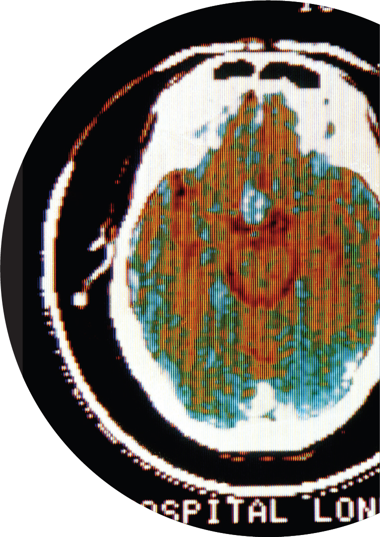
A woman with a suspected brain tumor received the first clinical computed tomography (CT) scan at Atkinson Morley Hospital in London on October 1, 1971. This non-invasive body imaging procedure relies on multiple X-ray transmissions, or “slices,” that are then reassembled by a computer to create 3D images of the body’s tissues and organs. Though early scanners took several minutes to create a single slice and days to create the reassembled image, modern CT machines take only a few seconds to generate images of the body.
1971
Advanced Drug Delivery Systems
A drug delivery system is a formulation or device that facilitates the introduction of a therapeutic substance into the body and improves its effectiveness and safety by controlling the rate, time, and place of release of drugs in the body. The first drug delivery systems with internal control of their rate of delivery of a therapeutic agent were developed in 1971. By the 1980s, advances in biotechnology and molecular biology had led to systems that enabled ever greater control over the delivery of drugs to the body.
1974
The Origins of Magnetic Resonance Imaging
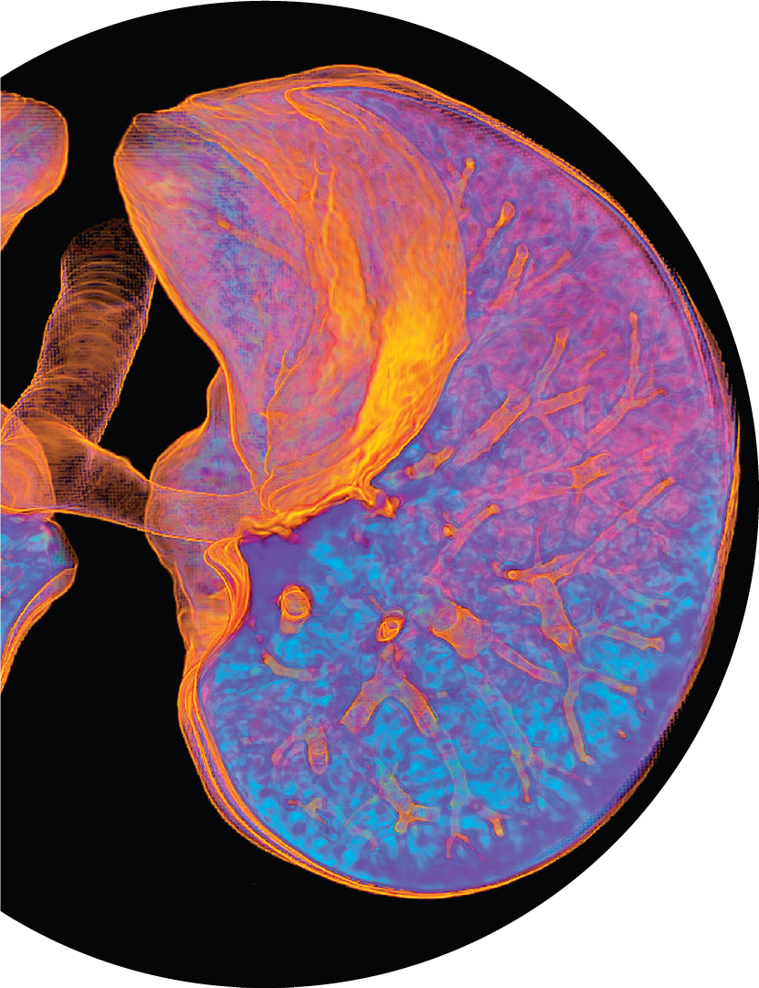
The quest to develop clinically useful applications of what would become known as magnetic resonance imaging (MRI) started after the first image was taken of a mouse using this technique in 1974. MRI scanners measure the speeds at which atoms in the body return to equilibrium after exposure to magnetic fields. The data then are reconstructed using computed tomography to create a 3D image of the body’s internal tissues, including tumors or tissue damage.
1974
Increasingly Specific and Sensitive Imaging
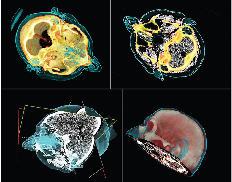
Positron emission tomography (PET) generates images by detecting the energy given off by decaying radioactive isotopes following their injection into the body. First developed in 1974, PET scanning is the most specific and sensitive method to image molecular interactions and pathways within the human body, using biomarkers to reveal information about disease, pharmaceutical effects, and many other biological processes. Hybrid PET/CT and PET/MRI imaging systems are now common in clinical settings.
1978
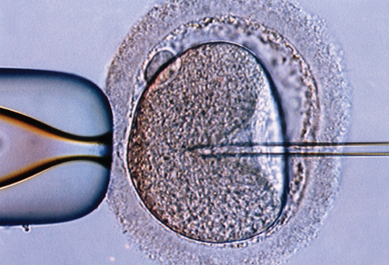
The First Baby Conceived in a Laboratory
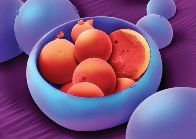
The world’s first “test tube baby” conceived via in vitro fertilization (IVF) was born on July 25, 1978, in Aberdeen, Scotland. In this procedure, mature eggs retrieved from the mother’s ovaries are fertilized with the father’s sperm in a laboratory dish and incubated briefly before the embryo is implanted into the mother’s womb. Since 1978, millions of children have been born using IVF, which is now viewed as a mainstream medical treatment for infertility. However, the procedure remains far from perfect, with fewer than one-third of attempts ending in a live birth.
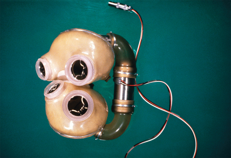
1982
The First Artificial Heart
In 1982, a dentist received the first totally artificial heart to permanently replace his failing natural heart. This device, called the Jarvik-7, featured a pump that replicated the lower two chambers of the heart, providing blood flow to the rest of the body. Though the operation was a success and the patient lived on the Jarvik-7 for 112 days, his quality of life was poor, tethered to a large air compressor and suffering from convulsions, kidney failure, and memory lapses before his death. Subsequent patients with artificial hearts fared better, but work on the “holy grail” of modern medicine, a permanent synthetic heart, continues today.
1984
A Bridge to Heart Transplantation
In 1984, the left ventricular assist device (LVAD) made history when it began keeping heart patients alive until donor hearts became available for transplantation. A shortage of donors has led to improvements to these devices that have enabled them to be used as long-term alternatives to heart transplants.
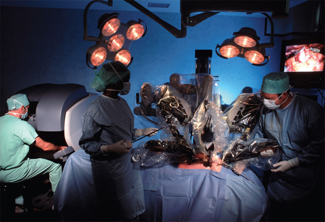
1985
Robotic Assistance in Surgery
In 1985, surgeons used a robotic arm for the first time to perform a delicate brain biopsy. Since then, robotic systems have become increasingly adept, generally in combination with flexible fiber optic cameras. With their steadily improving dexterity, accuracy, and visualization, robotic systems can now perform minimally invasive procedures that result in shorter hospital stays and quicker recovery times.
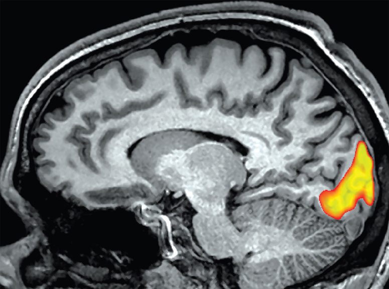
1992
Watching the Brain Function
MRI is so powerful that it can detect changes in the oxygenation level of the blood. In 1992, several groups began using this technique to map activity in the human brain. Functional MRI (fMRI) imaging can be used to study any motor, sensory, or cognitive task that a patient can perform while in a scanner. Applied clinically to preserve critical functions in patients needing neurosurgery, it has produced groundbreaking insights into how the brain functions in health and disease.
2008
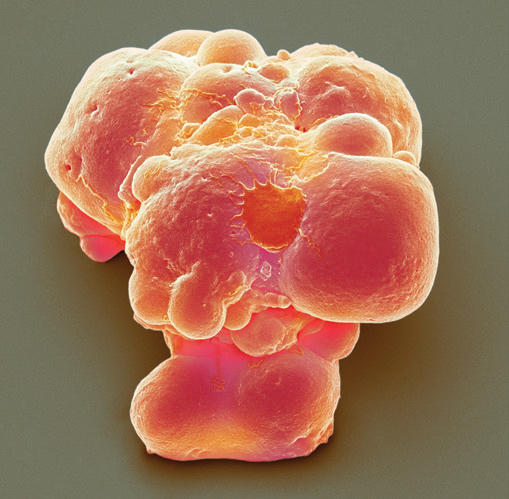
Reprogramming Human Body Cells
Almost all of the cells in the human body have developed into a specific type of cell and cannot change—they are skin cells, heart cells, brain cells, and so on. But in 2008 scientists learned how to reprogram human body cells through a process called induction, converting them into cells with the potential to develop into many different cell types. These induced pluripotent stem cells have become vital tools for research and drug development and may someday be used to generate tissues and organs for transplantation.
2020
RNA Vaccines
A classical vaccine works by artificially introducing a weakened or inactivated infectious agent (called an antigen) into the body, stimulating the immune system to produce antibodies that will fight against that infectious agent in the future. In contrast, RNA-based vaccines contain genetic instructions that the body uses to produce antigens, which trigger the immune response to create disease-specific antibodies. The development and use in 2020 of several RNA-based vaccines that effectively protect against COVID-19 will likely drive huge growth of this technology. In the future, RNA vaccines may allow for a single vaccination to protect against multiple diseases.

As the quest for better medical imaging devices, diagnostic measures, and therapeutic tools advances, health care will continue to improve. Diagnoses will become more accurate, procedures less invasive, and treatments more effective.


