
The integration of information from numerous types of biomarkers provides a clearer picture about a person’s traumatic brain injury (TBI), said Luca Marinelli, GE Research. This chapter highlights speaker presentations that focused on the state of the science in four major classes of TBI biomarkers, their current levels of evidence, and where further research is needed.
CLASSES OF TBI BIOMARKERS
Major classes of TBI biomarkers include those identified through neuroimaging, proteins detectable in blood and other biological fluids, electrophysiological signals, and other physiological indicators such as those measured through eye tracking or gait analysis. This section describes the landscape of current knowledge on the development and use of these types of biomarkers in TBI care and research.
Neuroimaging Markers1
Elisabeth Wilde, University of Utah, reviewed data on several prominent imaging techniques that can be used as biomarkers of brain injury, particularly computerized tomography (CT) and magnetic resonance imaging (MRI). These are both noninvasive options for imaging the brain. CT remains an important method in clinical guidelines for TBI, although it has
___________________
1 This section is based on a presentation by Elisabeth Wilde, University of Utah.
sensitivity limitations in cases of acute mild TBI and chronic TBI. CT imaging is less costly and generally faster than MRI and can be a better option in trauma emergencies. However, MRI does not use ionizing radiation as used in CT, and radiation exposures can be a concern for younger children or when multiple imaging exams are needed. MRI also provides greater detail in visualizing abnormalities within the brain.2 Neuroimaging markers are best established for acute TBI that is moderate and severe in nature. Progress is being made in developing more advanced imaging biomarkers, including a variety of MRI markers, although many do not yet reach level 1 evidence and require additional validation.3 However, Wilde noted that the literature on imaging for TBI is far ahead of clinical practice, reflecting the importance of clinical implementation discussed in the prior session.
There has been a dramatic increase in TBI imaging publications since 2015, with T1-weighted MRI and diffusion MRI being two modalities growing the fastest. Different imaging modalities are related to the types of pathology they can identify, and use of different modalities will be necessary to capture the heterogeneous pathology that characterizes TBI. For example, diffusion imaging and susceptibility-weighted MRI imaging are most commonly used to look for vascular changes, metabolic alteration is typically assessed using spectroscopy, and positron emission tomography (PET) can be used to detect neuroinflammation.
Diffusion and volumetric analysis provide insight into structural changes in the brain postinjury and are promising as diagnostic TBI markers. Even in mild TBI, studies have reported abnormalities in diffusion metrics. However, the limited longitudinal studies so far conducted do not suggest a consistent pattern of diffusion change over time. Because of the cost of MRI, Wilde explained, imaging is generally taken at several points rather than over multiple, short intervals, and it is difficult to have a precise understanding of the trajectory. Additional questions in using imaging for TBI diagnosis include which modalities to measure (what), the optimal time points to use (when), and which brain structures to visualize (where), given the diversity of injuries. She also noted the difficulty of validating imaging markers in mild TBI owing to inconsistent diagnostic criteria for this category of injury.
___________________
2 See https://radiology.ucsf.edu/blog/neuroradiology/exploring-the-brain-is-ct-or-mri-better-for-brain-imaging (accessed December 15, 2022).
3 The strength of evidence in analyses of the literature and development of recommendations can be categorized by different levels, based on how the evidence has been obtained (for example, whether it is drawn from analysis of one or more well-designed randomized controlled trials [RCTs] versus derived from cohort studies, expert opinion, or other sources). Level 1 evidence is considered the strongest and generally drawn from systematic reviews or from several RCTs with consistent results.
Neuroimaging markers may also be useful as prognostic and predictive biomarkers. Most studies in adults have found a relationship between diffusion imaging results and symptom presentation, but establishing this relationship remains tenuous in children. However, Wilde noted a recent pilot study using diffusion imaging and machine learning algorithms to predict symptoms after mild TBI (such as concussion) in children. The method was able to predict recovery, but with low sensitivity and specificity (Fleck et al., 2021). Another study in children using diffusion imaging and machine learning showed increased diagnostic accuracy (Mayer et al., 2022), but these techniques are not sufficiently advanced to implement in clinical practice.
The use of neuroimaging to monitor injury progression is the area that has seen the least investigation so far, she reported, although some studies have explored brain effects in older adults with a prior history of TBI as well as the alteration of brain development after injury in children.
Challenges for the clinical implementation of imaging biomarkers include the pace of evolution of the field—particularly upgrades in scanner hardware and software that necessitate periodic reanalyses and recalibration of previously collected data so it can be compared across study sites and times. Balancing using the newest and most specialized imaging sequences versus those that are well validated and widely available across sites is also a challenge. A lack of consensus remains about common analytic pipelines or deployment of new methodologies using machine learning. One of the most significant challenges in neuroimaging, however, said Wilde, is the lack of normative data and the narrow indications for some existing datasets. Normative data is expensive to collect and has a limited shelf life because of hardware, software, and sequence developments, so data may be nearly obsolete by the time researchers are ready to analyze it. Available normative data is also commonly collected from college students and may not reflect sufficient demographic and social variables nor be representative of the full range of TBI patients.
Wilde concluded with recommendations building from a 2019 Brain Trauma Blueprint meeting convened by Cohen Veterans Bioscience (see Box 3-1). Implementing these recommendations involves efforts to develop common analytic pipelines and harmonize previously collected imaging data, use of consistent standards and calibration strategies, and the need for multimodal analysis to integrate information from multiple markers. Using TBI biomarkers for distinguishing TBI phenotypes for inclusion/exclusion criteria in clinical trials, for identifying which patients are most likely to benefit from specific interventions, and for monitoring and assessing the effects of an intervention also remain important directions.
Blood-Based Biomarkers4
Jessica Gill, Johns Hopkins University, discussed progress in the development of fluid biomarkers for TBI, focusing on proteins detectable in blood while noting that such markers can be studied in saliva, urine, and other bodily fluids. From prior studies it is known that levels of glial fibrillary acidic protein (GFAP) and ubiquitin C-terminal hydrolase L1 (UCH-L1) are related during the acute period of experiencing a TBI, while neurofilament light chain (NfL) and Tau proteins are associated with recovery over the post-acute period. She highlighted several areas of study that are advancing the identification and measurement of blood biomarkers to provide the most reliable signals, noting the similarity of issues to those also being investigated in the neuroimaging field. Areas she highlighted include efforts to do the following:
- Identify novel biomarkers to increase diagnostic and prognostic sensitivity and specificity (moving beyond well-studied markers such as GFAP and UCH-L1), including strategies to measure proteins found in low quantities.
- Map objective fluid and nonfluid (imaging) biomarkers to study the specific pathologies and areas of the brain that have been affected after a TBI.
- Understand the clearance of biomarker proteins from the brain to blood, including patterns associated with different injury mechanisms (blunt force trauma versus blast injury) and in relation to factors that affect the types and concentrations of proteins measured in the blood, including time of day, nutritional status, and hydration.
- Study exosomes to identify the cellular origins of biomarker proteins, and study communication between cells.
- Investigate the roles of individual variability associated with clinical and demographic factors, including underlying, more complex social-environmental factors that shape exposure risk and recovery and that are just starting to be understood.
Gill shared data from the CARE Consortium,5 which includes baseline data on protein levels and gene activities measured before an individual plays a collegiate sport, facilitating comparison after concussion. She high-
___________________
4 This section is based on a presentation by Jessica Gill, Johns Hopkins University.
5 The Concussion Assessment, Research and Education (CARE) Consortium was established in 2014 with funding from the Department of Defense and National Collegiate Athletic Association (NCAA) to study mild traumatic brain injury in athletes and military cadets. See https://careconsortium.net (accessed December 15, 2022).
lighted a study in which they examined more than 8,000 proteins in blood collected from athletes at different time periods after concussion. The results identified the expression of erythrocyte membrane protein band 4.1 (EPB41) as upregulated and alpha-synuclein (SNCA) as downregulated after concussion. Having identified this combination, the investigators have been working on ways to measure it. Gill emphasized the technical importance of understanding protein isoforms (variants of a protein) and the need for careful testing and validation when developing and optimizing new biomarker assays, processes that can be time consuming. EPB4.1 is an abundant protein but has been more challenging to measure and validate, while the measurement of SNCA was being developed by Quanterix and can be measured and validated on their platform.
Using the CARE cohort, Gill also shared information from ongoing studies measuring blood biomarkers (GFAP, UCH-LI, NfL, and Tau) over 5 years in patients with different severities of TBI (Shahim et al., 2020a,b). Overall, the studied biomarkers map to volume loss changes seen in the brain over the first year, providing an indication that biomarkers can inform TBI risk and can be useful in monitoring.
Gill highlighted the opportunity to identify how fluid biomarkers relate to brain regions and forms of injury, which involves understanding the clearance of the proteins from the brain to the plasma. A study of 700 acute TBI patients used MRI and CT imaging and gadolinium contrast to look at brain meninges after injury and 3–5 days later to examine changes (Gill et al., 2018). Within this cohort, they found meningeal enhancement, in which there is a visual difference from normal in the appearance of the membrane surrounding the brain on imaging, to be the most predictive of long-term symptoms, with longer duration and greater magnitude of enhancement associated with more prolonged recovery. Meningeal enhancement is a difficult part of imaging, and it is not often studied, Gill noted, and it would be useful to have a biomarker that could indicate that a patient would benefit from including this assessment.
Biomarkers of inflammation have predictive value in TBI and relate to findings from brain imaging, but these inflammatory markers are not specific to neurotrauma. To better distinguish where inflammation is coming from, Gill’s team has developed protocols using exosomes, which are nanovesicles secreted by cells throughout the body. A major function of exosomes is in cell–cell communication—in which a nanovesicle secreted from one cell binds to and releases its contents into another cell, thereby affecting that cell’s function. Antibodies can be used to identify the cell type from which the exosome came, Gill explained, and exosomes detected in blood can be mapped back to brain cells such as microglia and astrocytes, adding a layer of spatial and functional information. As an example, she shared an early study in people in a particular military role (breachers) that results in signif-
icant blast exposures compared with unexposed controls. The findings were contradictory to prior studies that have found that being a military service-member or veteran increases inflammation overall. Instead, her team found that serum markers of inflammation measured in the studied breachers were lower than in controls. When using neuronally derived vesicles, however, inflammatory markers were significantly increased in the breacher group (Edwards et al., 2022), indicating differences specifically associated with the brain compartment. The findings suggest that exposure to high numbers of even low-level blasts can result in ongoing central inflammation, but that peripheral markers, such as serum concentrations, may be inadequate surrogates to detect this neuroinflammation.
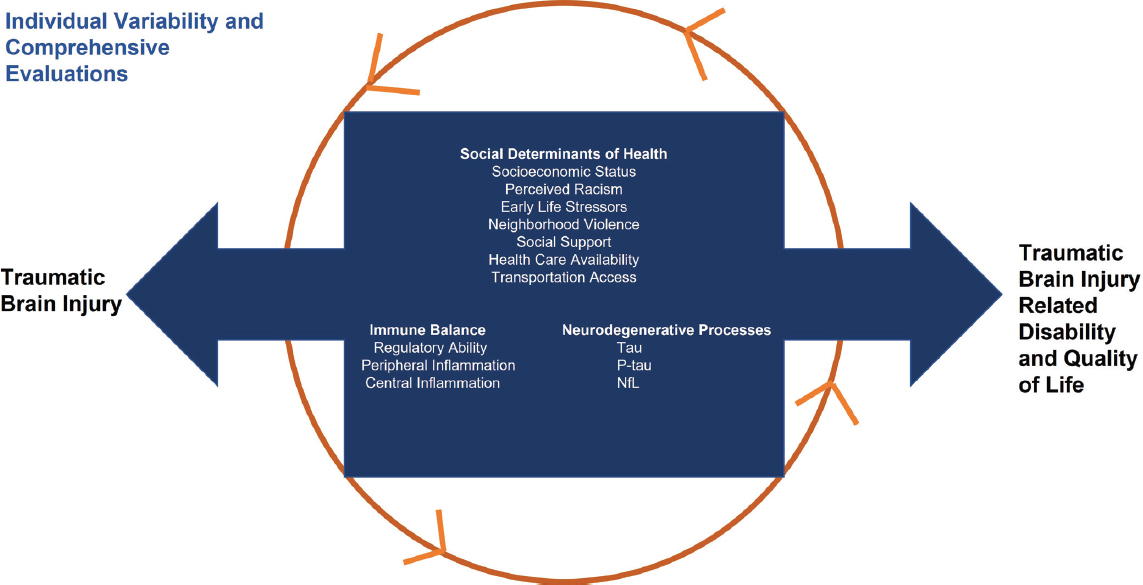
Putting all of this together, she said, identifying the right fluid biomarkers and matching them with imaging and other types of markers continues to be a challenge, but it is clear that something objective is happening in people with TBI that can be measured and translated into markers useful for different situations. Gill concluded with a call to rethink TBI not as an acute, discrete injury, but as a complex condition with multiple potential physiological and biochemical changes and neurological or psychological effects (see Figure 3-1). This can make it more difficult to recover from a TBI and perhaps places a person at further risk for additional TBIs.
Electrophysiological Markers6
Paul Rapp, Uniformed Services University and University of California, Irvine, described the advantages of using electroencephalograms (EEGs) in the assessment of TBI. EEG is also a noninvasive measurement technology. Rather than a static image, it provides information about electrical activity in the brain. EEG technologies are also portable and do not require sophisticated infrastructure, enabling them to be deployed in austere or military far-forward settings. EEG technologies provide immediate results and enable repeat EEGs to collect longitudinal data.7
More than half a dozen EEG-based biomarkers have been identified for TBI and have assays cleared by the Food and Drug Administration (FDA) for acute structural brain injury, acute functional impairment, and the presence of concussion. EEG is a readily usable technology for TBI,8 although Rapp recognized several challenges associated with measuring EEG and event-related potentials (ERPs). These measurements can be sensi-
___________________
6 This section is based on a presentation by Paul Rapp, Uniformed Services University and University of California, Irvine.
7 See, for example, Rapp et al., 2015a.
8 An EEG generally measures brain electrical activity continuously over a period of minutes, while an event-related potential (ERP) is a short segment of an EEG signal, showing the change in neuronal activity after a stimulus.
SOURCE: Presented by Jessica Gill, September 29, 2022.
tive to clinical factors that are difficult to control, such as circadian rhythm changes, recent exercise, fatigue, or drugs. They can also be sensitive to how they are collected, such as electrode positioning, and they are sensitive to electromagnetic interference. EEG and ERP measures are often used in conjunction with other forms of imaging or clinical examinations.
Taking a step back, Rapp reflected on the broader challenges applicable across types of TBI biomarkers, including the potential for a complex presentation and delayed clinical presentation. A single injury event can initiate more than one pathophysiological process, he said, and these can have different time courses and can interact with each other. A person’s biological response to an event is also dependent on past exposures and history of TBI. Further, people can present in a clinic when their injuries were sustained months or years prior, but their symptoms have reemerged.
In response to these challenges, Rapp shared examples of actions being taken by the field. Work is being done on expanding the mathematical approaches to analyzing EEG data, he said, including use of interregional synchronization and the development of time-dependent measures of interregional information movement, such as time-dependent transfer entropy. Network analysis—a type of statistical analysis that identifies relationships between items—has also become a major area of study and is being applied to the understanding of information handling in the central nervous system. Lastly, methods are being employed to look at signal changes, including using quadrant scanning of recurrence diagrams to detect changes that cannot be detected by conventional means.
In addition to expanding analysis techniques, efforts to expand data acquisition from EEGs and ERPs are ongoing, as are expanded analyses to incorporate additional metrics for multivariate discrimination. An indicator such as heart rate variability (HRV) can be easily tracked by a wearable device and combined with data from genomics and transcriptomics, eye tracking, and individual clinical history and analyzed using statistical learning technologies to determine which of the measures are most diagnostically or prognostically useful.
An example of a prototype that incorporates diverse information is the Integrated Neuropsychiatric Assessment System, which includes an electrode cap, digital amplifiers, tachistoscope, and laptop, all designed for use in rugged environments (Rapp et al., 2015b). The system is designed to handle the simultaneous acquisition of EEGs, ERPs, and eye tracking, information that can be combined with patient history, imaging and protein biomarker data, and psychological assessments to produce an integrated analysis. Rapp concluded that collecting multiple types of signals and using mathematical analysis approaches will advance the ability to bring information from multiple types of markers and assessments together in a clinically usable fashion. A recent review of psychophysiological biomarkers, such as the integration
of data from HRV, EEG, and other psychological and behavioral measures, identifies ongoing challenges and cautionary observations for the field as it continues to advance (Rapp et al., 2022).
Other Physiological Markers for TBI: Thinking Outside the Biomarker Box9
Other types of markers can be measured to detect clinically observable phenomena after TBI, said Christina Master, Children’s Hospital of Philadelphia. These measures may have potential for improving the understanding of subclinical physiological dysfunction, as well, particularly in areas with functional implications for patients in their everyday lives, such as visual and vestibular issues. Information from these other types of markers may also identify potential targets for interventions aimed at helping patients return to full function.
Looking across the spectrum of injury, mild TBI ends up being one of the most challenging scenarios for the use of biomarkers, she said. For many decades, less serious forms of TBI were often written off as insignificant because physiological signs of the symptoms that patients were describing were not observable and deficits were too subtle to measure. As with other forms of TBI, the goal of identifying and using biomarkers for milder injuries is to help diagnose, prognose, treat, monitor, and return patients to full recovery and function.
Master described potential targets for measurement. Autonomic dysfunction is one such area, and applying exercise physiology to TBI has demonstrated exercise intolerance after concussion. A measure such as heart rate threshold on the Buffalo Concussion Treadmill Test, which is the heart rate at which concussion symptoms are provoked, is prognostic, and patients who could not elevate their heart rate to 135 beats per minute were more likely to have prolonged recovery after TBI (Leddy et al., 2018). Prescribing aerobic exercise has been found to be therapeutic and to aid in preventing persistent postconcussion symptoms (Leddy et al., 2021). There is additional interest in using HRV as a biomarker, as Rapp mentioned. While more research is needed, HRV is a potentially prognostic biomarker. It is also sensitive to a history of concussion and may reflect residual effects from TBI even after apparent clinical recovery (Harrison et al., 2021).
Eye tracking is another active area of research, with some devices demonstrating the ability to distinguish between people with and without concussion (Bin Zahid et al., 2020). After brain injury, a common symptom is convergence insufficiency, in which the eyes do not coordinate binocularly
___________________
9 This section is based on a presentation by Christina Master, Children’s Hospital of Philadelphia.
for tracking objects.10 Eye measurement modalities include infrared eye tracking, retinal image-based tracking scanning laser ophthalmoscopes, and measuring smooth pursuit eye movements, with some of these devices achieving FDA clearance as an aid to concussion diagnosis, such as EyeBox (Bin Zahid et al., 2020) and Eye Sync (Sundaram et al., 2019). Such technologies may potentially be used in the future on the sidelines of a playing field to inform whether players can return to play following an injury. Objective tracking may also be used in the future to measure outcomes in a trial of occupational therapy for concussion-related convergence insufficiency.
From eye tracking, Master moved to vestibular coordination, where eye tracking also plays an important role, and she said that companies and researchers are looking at both motion sensitivity and motion issues such as balance and gait. Reduced saccade velocity and changes in smooth pursuit eye movement have been associated with the decreased ability of the eyes to track a moving field (optokinetic nystagmus) after concussion (Kelly et al., 2019).11 Postural control and gait are other areas of study within vestibular coordination, and research has found that center-of-mass range of motion and lateral step variability are associated with persistent postconcussive symptoms (Howell et al., 2020). The ability to capture these physiological measures in real-world settings, including through devices such as wearable sensors, is valuable. More work in this area is needed, however, Master emphasized, as self-reported dizziness in one study did not correlate with objective measures (Smulligan et al., 2021). The experience of mild TBI is still incompletely understood, and current physiological measures may not yet be fully capturing the symptoms experienced by patients.
Another potential visual biomarker, pupillary light reflex (PLR), may be relevant across the spectrum of injury severity, Master said. PLR lends itself to quantitative measurements and to monitoring over time. PLR metrics, in combination with standard demographic factors and CT findings, have been shown to be predictive of poor 6-month outcome in severe TBI cases (Romagnosi et al., 2022), and can be used to distinguish healthy adolescents from those with concussion (Master et al., 2020). This marker has the sensitivity to detect subclinical effects and could be used early on to see who might benefit from interventions and how to direct resources (Joseph et al., 2019).
___________________
10 https://www.nei.nih.gov/learn-about-eye-health/eye-conditions-and-diseases/convergence-insufficiency (accessed December 20, 2022).
11 Both saccade and smooth pursuit are types of eye movements, with the key point being that traumatic brain injury is associated with changes in the ability of a patient to perform certain visual functions. As defined in Purves et al., “Saccades are rapid, ballistic movements of the eyes that abruptly change the point of fixation,” while “smooth pursuit movements are much slower tracking movements of the eyes” (Purves et al., 2001).
Moving to auditory biomarkers, Master explained there is interesting work being done in areas such as the frequency-following response evoked by speech and the implications for TBI diagnosis, prognostication, and clinical care. Even after concussion, a diminished speech-evoked response has been observed, reflecting poor neural processing of sound. This response improves over time as other symptoms improve, showing its potential use in monitoring recovery. A signature of diminished response remains even after apparent clinical recovery, and so the marker is also sensitive to history of a prior concussion (Kraus et al., 2016, 2017).
Lastly, she highlighted altered sleep after concussion, using actigraphy to quantify how sleep is affected. Sleep quality tends to recover over 4 weeks. However, studies have found poor correlation between self-reported symptoms in those with sport-related concussion and objective measures captured through actigraphy (Considine et al., 2021). This demonstrates that there is still a need to improve understanding of altered physiology after traumatic brain injury and a need to find ways to close the gap between what can be measured and what patients report experiencing. Collecting observable physiological measures through wearables, conducting ecological momentary assessments in real time and real-life contexts, and incorporating options for remote patient monitoring can all be brought to bear in measuring and monitoring outcomes that are meaningful for patients.
DISCUSSION
Initial discussion centered around the interpretation and generalizability of biomarker results, given small sample sizes used in some studies, and the resulting implications for biomarker translation into clinical practice. From a clinician perspective, Master said that new biomarkers need to provide useful information as well as being feasible and user-friendly to be adopted in practice. Rapp indicated that some biomarker results can be overinterpreted and called attention to the importance of considering the confidence intervals when interpreting study results, saying that the intervals need to be tightened to be useful and that failures to replicate biomarker results can discourage clinicians from using them. Wilde added that the numbers of study participants are much larger in the work now being done through the growing number of TBI research consortia.12 In the
___________________
12 Examples of TBI research consortia mentioned during the workshop and recent report (NASEM, 2022) include the TBI Model Systems Network (https://msktc.org/about-modelsystems/TBI); Transforming Research and Clinical Knowledge in TBI (TRACK-TBI; https://tracktbi.ucsf.edu); CENTER-TBI (https://www.center-tbi.eu); NCAA-DoD Concussion Assessment, Research and Education (CARE) Consortium (https://careconsortium.net); and the Long-Term Impact of Military-Relevant Brain Injury Consortium and the Chronic Effects of Neurotrauma Consortium (LIMIC-CENC; https://www.limbic-cenc.org), among others.
last 5 years, she said, investigators have been able to collect large amounts of data that did not previously exist, and this has been of major benefit to the field. Several participants also noted the continued role for increasing understanding on when and how biomarkers can be used to inform TBI diagnosis and classification, prognosis, and recovery monitoring.
Kathy Lee, Department of Defense (DoD), commented that each quarter DoD receives a briefing on FDA-cleared biomarker devices through a DoD community of interest in TBI. There are currently eight cleared devices, she noted, but DoD is using very few of these yet. While biomarker devices and their cleared uses continue to evolve, she surmised that if a device is not yet usable in a point-of-care battlefield or forward military setting, some military clinicians may assume it is not yet useful in other types of care settings. It is also important, she said, for the biomarker field not to focus only on specialty services for brain injury but also to solicit feedback that informs the development of markers and assays usable in general locations, such as by emergency medical services and in hospital emergency departments. Master noted that in a primary care setting, providers will feel most comfortable using their existing, familiar clinical assessment tools. While new technologies and modalities will inform a more refined characterization of TBI, not all technologies may become a standard part of clinical practice.
Another participant asked how to let the data teach researchers and clinicians what the most valuable biomarkers are, instead of trying to identify every possible marker. Gill replied that lessons can be gleaned from genomics, where researchers look across the genome to identify candidate genes, analyze their relationships in gene expression networks, and validate predictive value. Now that technologies enable many simultaneous measurements, she said, researchers can use such network analyses of biomarker proteins to establish profiles characteristic of different subtypes of TBI. Her lab has used these approaches to collect pilot data, subsequently seeking funding to validate the data in different and expanded cohorts. She called for discovery platforms within TBI consortia that are additive and flexible, and for grant funding to enable findings to be built on as they emerge, instead of discrete silos for predetermined topics.
Frederick Korley, University of Michigan, asked each speaker to select one biomarker ready for incorporation into a TBI classification system, particularly any with level 1 evidence as well as other markers in the pipeline that have level 2 or 3 evidence. In terms of fluid biomarkers, Gill said that NfL protein is performing well across consortia for both acute and chronic TBI patients. Phosphorylated tau has been advancing as a prognostic biomarker, as well, she added. For imaging, Wilde said that diffusion imaging does not yet reach level 1 but is the most promising and widely used MRI modality at this point. Master acknowledged that evidence for the categories of physiological markers she discussed are behind imaging
and fluid biomarkers. It will be years before they are ready for clinical use, she noted, but they can be useful in distinguishing subtypes of TBI. Lastly, Rapp suggested that EEG in conjunction with neuropsychological testing can serve a useful combination marker.
This page intentionally left blank.


