National Academies Press: OpenBook

Radioactive Sources: Applications and Alternative Technologies (2021)

Chapter: Appendix F: Sterilization Using Radiation with Different Modalities

« Previous: Appendix E: Economic Feasibility of Adopting Alternative Technologies
Suggested Citation:"Appendix F: Sterilization Using Radiation with Different Modalities." National Academies of Sciences, Engineering, and Medicine. 2021. Radioactive Sources: Applications and Alternative Technologies. Washington, DC: The National Academies Press. doi: 10.17226/26121.
×

Appendix F

Sterilization Using Radiation with Different Modalities

The process of sterilization using gamma ray, x-ray, and electron beam (e-beam) is broadly similar in terms of the transfer of energy and interaction. Gamma rays and x-rays both involve a two-step process of photons interacting with the material, primarily through the Compton effect, and then the created secondary electrons deposit the dose and perform the damage to the biological structure of the bioburden. In the case of e-beam, the initial interaction of photons with the material is omitted, and instead, electrons interact directly with the material. That is, e-beam is the most direct modality of sterilization. This difference between e-beam and gamma or x-ray sources is that it leads to different dose distributions in the product.

X-rays have slightly better penetration than gamma rays from cobalt-60 (see Figure F.1). As with gamma rays, x-rays generate electrons, which are the active factor when interacting with the product to be sterilized. The bremsstrahlung process generates x-rays that are generated for the purpose of sterilization. Typically, 7.5-MeV electrons are directed onto a dense, high-Z material such as tantalum. As the electrons are scattered by the target material atoms, a broad spectrum of x-rays is produced (see Figure F.2). The bremsstrahlung process is very inefficient at 7.5 MeV; only 10–15 percent of the electron energy is converted to x-rays. The rest is dissipated as heat in the target. Therefore, to generate 15 kW of x-ray power, approximately 120 kW of electron beam power is required. A megacurie of cobalt-60 releases approximately 15 kW of photon energy.

E-beam accelerators used in sterilization are typically in the 50- to 80-kW range, which is equivalent to 3 to 5 MCi (111 to 185 GBq) of cobalt-60. The dose rate of an e-beam can be as high as 20 MGy/hr allowing the processing of a box or carton of product within tens of seconds. The throughput of an e-beam facility can be designed to be comparable to that of a gamma facility.

One parameter that may cause a difference in the response of a material to the three radiation modalities for sterilization is the dose rate. A gamma facility typically delivers approximately 10 kGy/hr. For a typical sterilization prescription (25 kGy) this means that a device must remain in the irradiation chamber for 2.5–3 hours. The dose rate for an x-ray system can be six times the rate of gamma, resulting in an irradiation time of 20–30 minutes. E-beams can deliver approximately 20 MGy/hr, and products here can be irradiated in seconds. This dose rate variation can be advantageous or disadvantageous depending on the material that is being sterilized. Some undesired reactions in materials may not have time to develop at higher dose rates, improving their ability to tolerate irradiation. However, 25 kGy in water causes a 6°C increase in temperature. This could be higher in certain areas of a product depending on variations in density and other geometrical factors. A sharp temperature increase in a short period of time could be a concern or it could be more tolerable than spending 2–3 hours in the elevated temperature environment of a typical cobalt-60 irradiator.

Suggested Citation:"Appendix F: Sterilization Using Radiation with Different Modalities." National Academies of Sciences, Engineering, and Medicine. 2021. Radioactive Sources: Applications and Alternative Technologies. Washington, DC: The National Academies Press. doi: 10.17226/26121.
×
Image
FIGURE F.1 Plot of penetration depth of the three sterilization radiation modalities.
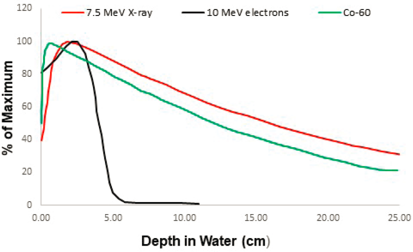
NOTES: Data are for water (1 g/cm3). X-ray data are for 7.5 MeV electrons on a bremsstrahlung target.
SOURCE: Fermi National Accelerator Laboratory.
Image
FIGURE F.2 Energy spectra of the three sterilization radiation modalities.
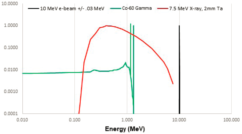
NOTES: The electron beam curve includes a 30-keV width, which is readily achievable by accelerator sources. The spectrum from that was then attenuated by 2 mm of tantalum to represent self-absorption in the bremsstrahlung target.
SOURCE: Fermi National Accelerator Laboratory.
Suggested Citation:"Appendix F: Sterilization Using Radiation with Different Modalities." National Academies of Sciences, Engineering, and Medicine. 2021. Radioactive Sources: Applications and Alternative Technologies. Washington, DC: The National Academies Press. doi: 10.17226/26121.
×
Page 181
Suggested Citation:"Appendix F: Sterilization Using Radiation with Different Modalities." National Academies of Sciences, Engineering, and Medicine. 2021. Radioactive Sources: Applications and Alternative Technologies. Washington, DC: The National Academies Press. doi: 10.17226/26121.
×
Page 182
Radioactive Sources: Applications and Alternative Technologies Get This Book
×
Buy Paperback | $75.00 Buy Ebook | $59.99
MyNAP members save 10% online.
Login or Register to save!
Download Free PDF

Radioactive Sources: Applications and Alternative Technologies assesses the status of medical, research, sterilization, and other commercial applications of radioactive sources and alternative (nonradioisotopic) technologies in the United States and internationally. Focusing on Category 1, 2, and 3 sources, this report reviews the current state of these sources by application and reviews the current state of existing technologies on the market or under development that are or could be used to replace radioisotopic technologies in those applications. Radioactive Sources will support existing and future activities under the National Nuclear Security Administration Office of Radiological Security program to reduce the use of high-risk radiological materials in commercial applications.

  1. ×

    Welcome to OpenBook!

    You're looking at OpenBook, NAP.edu's online reading room since 1999. Based on feedback from you, our users, we've made some improvements that make it easier than ever to read thousands of publications on our website.

    Do you want to take a quick tour of the OpenBook's features?

    No Thanks Take a Tour »
  2. ×

    Show this book's table of contents, where you can jump to any chapter by name.

    « Back Next »
  3. ×

    ...or use these buttons to go back to the previous chapter or skip to the next one.

    « Back Next »
  4. ×

    Jump up to the previous page or down to the next one. Also, you can type in a page number and press Enter to go directly to that page in the book.

    « Back Next »
  5. ×

    Switch between the Original Pages, where you can read the report as it appeared in print, and Text Pages for the web version, where you can highlight and search the text.

    « Back Next »
  6. ×

    To search the entire text of this book, type in your search term here and press Enter.

    « Back Next »
  7. ×

    Share a link to this book page on your preferred social network or via email.

    « Back Next »
  8. ×

    View our suggested citation for this chapter.

    « Back Next »
  9. ×

    Ready to take your reading offline? Click here to buy this book in print or download it as a free PDF, if available.

    « Back Next »
Stay Connected!