
The fourth session of the workshop focused on efforts to modulate the host immune system to create a pro-regenerative environment. The session’s objectives were to (1) discuss the goal of host immune modulation and consider what the correct molecular targets are for creating a pro-regenerative environment and (2) examine recent advances of the role of innate and adaptive immunity in cell engraftment and endogenous tissue regeneration, and approaches for immunomodulation of the structure and function of stem cell niches for goals of tissue regeneration. The session was moderated by Candace Kerr, program officer of the Stem Cell Program of the Aging Physiology Branch at the National Institute on Aging.
CELLULAR SENESCENCE, SENOLYTICS, AND ORGAN REGENERATION AND TRANSPLANTATION
James Kirkland, director of the Robert and Arlene Kogod Center on Aging and Noaber Foundation Professor of Aging Research at the Mayo Clinic, discussed fundamental aging processes and their effects on issues related to organ regeneration and transplantation.
Unitary Theory of Fundamental Aging Mechanisms
Fundamental aging processes begin at the time of conception, Kirkland stated. These processes, which are largely conserved across vertebrate species, can be classified into between four and 13 different categories or “pillars” of aging. Increasing evidence that these processes are interlinked, such that targeting one tends to affect the rest, underpins the unitary theory of fundamental aging processes (see Box 6-1), he explained. In addition, the geroscience hypothesis put forth by Gordon Lithgow holds that interventions modifying aging biology can slow its progression, thereby delaying or preventing the onset of multiple diseases and disorders (Sierra et al., 2021). Moreover, fundamental aging processes may be root-cause contributors to the majority of conditions that cause most morbidity, mortality, and health expenditures (see Box 6-1). In addition to aging phenotypes and geriatric syndromes (e.g., sarcopenia, frailty), these conditions include major chronic diseases—many of which still lack effective treatments—as well as decreased ability to withstand an infection, respond to a vaccine, or to recover after chemotherapy. Importantly, interventions that target any one of these fundamental aging processes will tend to affect all the others, Kirkland emphasized.
Transient, Beneficial versus Persistent, Deleterious Senescent Cells
Senescent cells accumulate not only with aging, but also at sites of age-related diseases, diseases in younger individuals, and multiple acute and chronic diseases, Kirkland explained. Senescent cells can be sorted into two groups: those that are acutely generated and beneficial versus those that are persistent and deleterious, he described. Newly formed senescent cells have lost the ability to divide but remain alive and can have myriad beneficial functions that include secreting a range of cytokines, chemokines, proteases, growth factors, noncoding nucleotides, and bioactive lipids. Furthermore, acutely generated senescent cells defend against cancer development and infection and support wound healing and tissue remodeling. Even in seemingly unrelated processes like parturition, senescent cells accumulate in the placenta and produce factors that drive the neonate through the birth canal. Since many physiological functions depend on the acute generation of transient senescent cells, Kirkland cautioned against interfering with the capacity of such cells to become senescent, which could lead to cancer development or failed wound healing. Although many or most senescent cells can produce senescent-associated secretory phenotype (SASP) factors, approximately 30–70 percent of persistent senescent cells may have a tissue-destructive, pro-apoptotic SASP, which appears to contribute to a host of deleterious effects and accentuate the other pillars of aging (Tripathi et al.,
2021). For example, persistent, SASP-expressing, pro-apoptotic senescent cells can result in (1) increased inflammation, fibrosis, and stem and progenitor cell dysfunction; (2) decreased levels of proteins such as nicotinamide adenine dinucleotide and α-Klotho; (3) elevated CD38 and mammalian target of rapamycin (mTOR); (4) increased likelihood of cancers and chronic disease; and (5) the spread of senescence.
Effects of Transplanting Senescent Cells
Murine models have been used to study the effects of transplanted senescent cells. For instance, transplanting relatively small numbers of senescent cells into a middle-aged mouse can cause physical dysfunction and result in premature death, Kirkland described (Xu et al., 2018). Transplanting 1 million senescent cells—such that scarcely one in 10,000 cells in the mouse is a transplanted senescent cell—is sufficient to drive frailty in those mice (Xu et al., 2018). The mice exhibit decreased strength, as shown by reduced hanging endurance and ability to run on a treadmill, and they tend to die early after a lag period. Mice with transplanted senescent cells tend to die prematurely due to all typical age-related diseases, not any specific disease, Kirkland highlighted. These findings support a causal link between senescent cells and multimorbidity, since adding a small number of senescent cells can accelerate age-related health problems and death from all causes (Xu et al., 2018).
Evidence also indicates that transplanted senescent cells can spread senescence to non-senescent host cells in both paracrine and endocrine manners, said Kirkland. By labeling senescent cells that were transplanted into a middle-aged mouse, researchers determined whether observed senescent cells originated from the transplanted senescent cells or if the recipients’ own cells became senescent (Xu et al., 2018). Remarkably, if senescent cells are transplanted into the peritoneum, the recipients’ cells in their arms and legs start becoming senescent, he said. The immune system normally removes senescent cells; however, these results suggest that, above a threshold burden, new senescent cells can be formed due to the spread of senescence at a rate that exceeds the ability of the immune system to clear them, Kirkland posited. When that threshold is surpassed, a range of deleterious effects may result. Experimental evidence from mouse studies further supports this postulated threshold phenomenon, he added. As previously described, approximately 1 million transplanted senescent cells are sufficient to cause dysfunction in a middle-aged mouse receiving a normal diet, but a transplant of only 500,000 cells has no injurious effect. Whereas, in old mice or middle-aged mice on a high-fat diet, a transplant of 500,000 cells can cause the recipient mouse to become frail and die prematurely (Xu et al., 2018). The outcome indicates that these mice are already closer to
the senescent-cell threshold burden due to the greater number of preexisting senescent cells in old or obese animals.
In another mouse study, transplantation of cardiac allografts from old donors also induced cellular senescence in young recipient organs, Kirkland observed. By comparing the effects of transplanting hearts from old mice into young mice versus from young mice into other young mice, researchers found that senescent cells from the hearts of the old mice spread senescence to other organs of the young mice (Iske et al., 2020). This occurs in much the same way as transplanting senescent cells into young mice, Kirkland commented. Furthermore, the 30–70 percent of senescent cells that have a pro-apoptotic, tissue-damaging SASP can secrete noncoding nucleotides, including mitochondrial DNA. Iske and colleagues showed that mitochondrial DNA produced by senescent cells in the hearts from the old mice activates dendritic cells in draining lymph nodes and substantially accelerates graft-versus-host disease (Iske et al., 2020). Kirkland noted that this effect is appreciated by transplant surgeons who are reluctant to transplant organs from old donors to young recipients, contributing to large numbers of nonutilized organs from consenting donors each year.
Hypothesis-Driven Senolytic Drug Development
For the past two decades, a new avenue of research has emphasized hypothesis-driven senolytic drug development, said Kirkland. In a seminal publication in 2004, Ned Sharpless and his colleagues found that caloric restriction, which increases healthspan, could reduce senescent cell accumulation as well as delay frailty and the onset of age-related diseases (Krishnamurthy et al., 2004). This work gave rise to questions about possible causal effects of eliminating senescent cells on morbidity and laid the groundwork for the lengthy process of developing drugs that would selectively target those senescent cells.
Given that the 30–70 percent of senescent cells with a tissue-destructive SASP survive despite killing the other cells around them, Kirkland and his team asked whether such senescent cells may have defenses against their own pro-apoptotic signaling. Through bioinformatics, an entire series of interlinked pathways emerged, forming a network termed the senescent cell anti-apoptotic pathway (SCAP) network. The networked anti-apoptotic regulator pathways confer resistance to apoptosis in senescent cells. Efforts to identify agents that target nodes in the network or along those pathways have yielded approximately 40–50 candidate drugs, Kirkland said. In particular, agents with short elimination half-lives are sought in order to kill or remove senescent cells in an intermittent “hit-and-run” fashion, given that it takes one to six weeks for new senescent cells to form in cell culture, he added. Since senolytic drugs target senescent cells rather than a single
molecule or pathway, the paradigm is analogous to developing antibiotics that could treat multiple types of infection, in contrast to the traditional one-drug, one-molecular-target, one-disease strategy, Kirkland highlighted.
Research into the mechanism of action of the senolytic agents has revealed that different types of senescent cells depend on different elements of the SCAP network for their survival, Kirkland stated. For instance, dasatinib is a Src kinase inhibitor that is senolytic for human fat cell progenitors but not for endothelial cells because human senescent fat cell progenitors rely on ephrin-dependent survival pathways to defend themselves against their own SASP. In contrast, endothelial-cell defense mechanisms depend more heavily on B-cell lymphoma 2 family members, p21-related pathways, and heat shock protein 90, so quercetin, a flavonoid, was effective against senescent human umbilical vein endothelial cells (HUVECs). Since dasatinib did not kill senescent HUVECs and quercetin did not kill senescent pre-adipocytes, a combination of the two agents was investigated in mice and was able to eliminate a broader range of senescent cell types than either agent could alone (Zhu et al., 2015). Furthermore, a comparison of transplanted luciferase-expressing senescent cells versus non-senescent cells showed that the 30–70 percent of senescent cells with a pro-apoptotic, tissue-damaging SASP were eliminated by allowing them to “commit suicide” based on their own SASP, Kirkland explained (Xu et al., 2018).
A series of other senolytics has been developed using these approaches and others, and the senolytics can delay, prevent, or alleviate more than 60 conditions in mice, Kirkland said. Although the translation of drugs from mouse to human is not straightforward, senolytics alleviated frailty when given to animals with transplanted senescent cells (Xu et al., 2018). In heart-transplanted animals, senolytics were associated with reduced rejection rates (Iske et al., 2020). Moreover, promising evidence suggests that treating the donor, the recipient, or even the heart itself with senolytics can potentially reduce rejection and death in mice, offering the potential to utilize greater numbers of older donor organs, remarked Kirkland.
However, it is not yet clear that the effects of senolytics in mice will safely translate to humans, noted Kirkland. To explore the safety and efficacy of these agents in humans, clinical trials of senolytics have already been initiated.1 Publications about the first-in-human clinical trials of senolytics have found that the agents can clear senescent cells effectively from human adipose tissue, for example (Hickson et al., 2019). Blood measures of senescence based on composite scores indicate that senolytic agents can reduce senescent cells through intermittent treatment. Within the Translational Geroscience Network, 15 clinical trials are underway to examine the effects of different senolytics on conditions such as frailty, Alzheimer’s disease,
___________________
1ClinicalTrials.gov identifier: NCT02848131.
diabetes-related kidney disease, idiopathic pulmonary fibrosis, osteoporosis, and osteoarthritis. One study is investigating the effect of senolytics after bone marrow transplantation in an attempt to delay the accelerated aging-like state that can develop three to five years after the transplant. Another study in childhood cancer survivors is using senolytics to attempt to reduce the incidence of the associated accelerated aging-like state. Three trials are also underway for coronavirus in inpatients, outpatients, and nursing home residents. A lot of work remains to be done to ensure safe translation of senolytic therapeutics to humans, Kirkland emphasized.
MAPPING THE IMMUNE AND TISSUE ENVIRONMENT IN HEALING AND NON-HEALING WOUNDS
Jennifer Elisseeff, Morton Goldberg Professor of Ophthalmology and Biomedical Engineering and director of the Translational Tissue Engineering Center at Johns Hopkins University, shared efforts to map the immune and tissue environment in healing and non-healing wounds.
Tissue and organ loss remains a global issue, said Elisseeff. Although transplantation can provide functional tissues for those suffering from tissue and organ loss, the major challenges of immune rejection and tolerance persist. Similarly, synthetic implants (e.g., cartilage, breast, and others) are associated with challenges because they do not recapitulate all tissue functions. These two fields and their limitations led to the development of tissue engineering, which aims to provide new healthy tissue by taking a biomaterial scaffold that serves as a three-dimensional framework for components like stem cells and where specified biological signals can promote tissue development. Unfortunately, clinical translation of tissue engineering discoveries has been slow, Elisseeff remarked.
Rebuilding Cartilage in Patients
In the early stages of tissue engineering research, Elisseeff and colleagues targeted cartilage tissue that lines the surface of joints because, when damaged, cartilage tissue cannot heal well. Elisseeff and colleagues began by designing materials to stimulate stem cell development and recapitulate elements of normal developmental biology to build replacement cartilage tissue, she explained. This work led to the development of therapies that combined synthetic and biological materials and could be incorporated with surgical practice to mobilize endogenous cells to promote tissue repair (Sharma et al., 2013; Wang et al., 2007). After 12 months, the surgical microfracture process leads to fibrocartilage that degrades over time;
however, tissue volume and quality can be maintained with a biomaterial scaffold (Sharma et al., 2013).
Clinical translation informed a new research direction when it became clear that this process did not recapitulate the elements of classical tissue engineering; instead, the treatment redirected the wound-healing process, Elisseeff said. Another clinical trial further demonstrated that there is a tissue-specific immune response associated with biomaterial implants (Hillel et al., 2011). Different types of immune cells and—unexpectedly—T cells were observed depending on which type of tissue was adjacent to the implant, she noted. Based on this discovery, their research focus shifted from classical tissue engineering to focus on the immune system as a key regulator of tissue regeneration and the biomaterial response. The use of biomaterials facilitated the development of models to study different tissue environments and how they mobilize the immune system, she added.
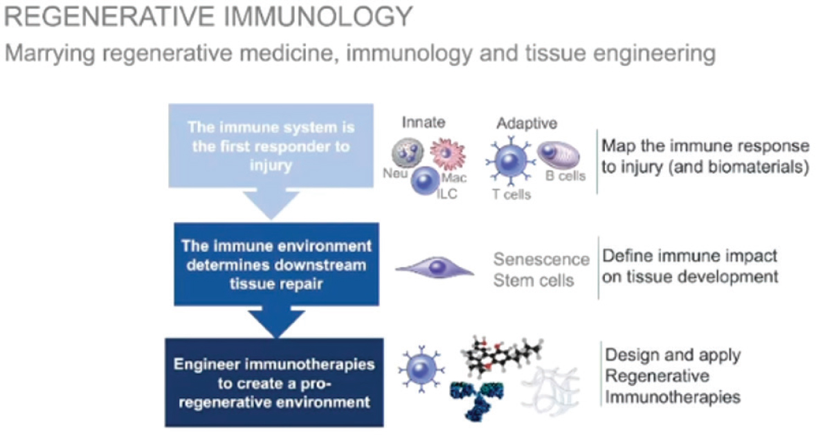
Regenerative Immunology
Elisseeff explained that the concept of regenerative immunology marries the fields of regenerative medicine, immunology, and tissue engineering (see Figure 6-1). As the first responder to an injury, the immune system sets the stage for subsequent healing processes. Although cells such as macrophages have long been recognized for their role in tissue repair, cells of the adaptive immune system, such as T cells, had not previously been considered as having a critical role. To inform efforts to engineer immunotherapies, Elisseeff and colleagues began mapping these immune responses and investigating how they influence downstream processes such as vascularization, tissue development, and mobilization of different immune cell types. The immune system is therapeutically accessible, making it a prime target for regenerative medicine, she noted.
Mapping the Natural Immune Response after Tissue Injury
A first step in developing immunotherapies to promote healing is to map the natural immune response after a tissue injury in a healing wound versus a non-healing wound characterized by fibrosis and inflammation, said Elisseeff. Muscle tissue is an example of a healing wound environment, and cartilage is a prototype of a non-healing wound, with biomaterials also modulating the immune environment.
Biomaterials Change the Immune Response
To examine the ability of biomaterials to modulate the immune response, Elisseeff and her team examined a pro-regenerative biological scaffold in a
SOURCE: Elisseeff presentation, November 3, 2021.
muscle wound and found that the biological scaffold stimulates interleukin 4 (IL-4) production by CD4 T cells and others as well as induces macrophage behavior to a pro-regenerative phenotype (Sadtler et al., 2016). Moreover, changes in both local and distal draining lymph nodes demonstrate how local wounds and implants can have systemic implications and, conversely, how the systemic state of the organism can influence a local wound’s environment, Elisseeff said. Some differences in the development of repair versus fibrosis depend exclusively on T cells, she added. If an animal without T cells is repopulated with healthy T cells, effective muscle repair can occur. However, if the animal receives T cells deficient in the mTOR complex 2, T helper 2 (Th2) cell differentiation cannot happen, leading to fibrosis and adipogenesis at the injury site (Sadtler et al., 2016).
Biomaterials-Directed Regenerative Immunology
In contrast to biologically derived biomaterials, synthetic materials are characterized by foreign-body-responsive fibrosis (Chung et al., 2020). For example, fibrosis and collagen are evident with clinical implants of a polyester-based scaffold, Elisseeff described. Whereas the biological scaffold stimulates IL-4, the synthetic material induces production of interleukin 17 (IL-17) by innate lymphocytes, gamma-delta T cells, and T helper 17 (Th17) cells (Chung et al., 2020). However, eliminating IL-17 with a neutralizing antibody can reduce that fibrosis, she noted. This finding is further supported with clinical data. In implants taken from patients and tested for the same markers, the production of elevated IL-17 by CD4 T cells and gamma-delta T cells exceeds the IL-4 interferon gamma production by these T cells around the clinical implants (Chung et al., 2020). The dichotomy of these biomaterial-specific immune responses can be thought of as a balance, Elisseeff described. The biomaterial can influence the environment toward a type 2 IL-4 response, with Th2 and eosinophils playing a major role and providing a healing environment, or the biomaterial can modulate toward a more fibrotic environment in which these cell types are secreting IL-17. This balance can be manipulated slightly by agents that are known to stimulate a type 2 response (e.g., Schistosoma soluble egg antigen) or inhibit IL-17, she added.
Regenerative Immunology and Big Data
Elisseeff and her colleagues are now integrating big data techniques, such as single-cell RNA sequencing, with the biomaterial models to create a large database of single-cell sequencing results from different biomaterial environments. To explore the communication modalities in the healthy versus fibrotic environments, researchers developed a program to examine
transcription factor activation in single-cell datasets and construct intercellular signaling networks, which can be grouped into modules (Cherry et al., 2021). Of particular interest is the communication among signaling modules that arise from the analysis, including an immune module, a fibroblast module, and a tissue module. Although some transcription factors and receptor combinations overlap in the different biomaterial and immune environments, others are unique. Elisseeff and her team are also using transfer learning algorithms to identify rare cell types, such as senescent cells, and can learn how the cells participate in immune–stromal communication.
Immunological Impact of Senescence
In non-healing cartilage wounds, the clearance of senescent cells reduces inflammation and improves tissue repair, Elisseeff commented. Senescent cells can be found in a non-healing cartilage wound characterized by inflammation and arthritis. Clearing senescent cells after injury can promote tissue repair simply by removing inhibitory factors without requiring additional growth factors or stem cells. However, senescent cells are immunologically active and impact multiple immune cell types, she noted. For example, the balance between IL-4 and IL-17 is particularly important for T cells, and the SASP of senescent cells at the injury site includes molecules known to influence them toward the less desirable Th17 pathway.
Further demonstrating this phenomenon, artificially induced senescent fibroblasts that were cultured with naïve T cells in the presence of TGF-beta induced a significant increase in IL-17 (Faust et al., 2020). Conversely, when T cells are guided toward the Th17 phenotype and cultured next to healthy fibroblasts, there is an increase in senescence markers. This phenomenon is referred to as “immunologically induced senescence.” In fact, a senescent cell may be considered an immune cell, with both IL-17 and senescent factors playing a role in a positive feed-forward loop, Elisseeff posited. In vivo experiments also provide evidence for immunologically induced senescence. Specifically, components such as gamma-delta T cells can increase IL-17 in the joint, and the lymph node exhibits associated changes. When senescent cells are removed, the IL-17 signature in the joint and lymph node decreases (Faust et al., 2020). In the context of the foreign body response, these findings present new therapeutic targets—namely, senescent cells and the immune factors that are capable of inducing senescence, such as IL-17, Elisseeff emphasized.
Aging Changes Immune Response to Injury
Aging alters wound repair and regeneration, Elisseeff said, noting that their procedure for local senolysis in the joint was not effective in older
animals, who required a combination of local and systemic senolysis (Faust et al., 2020). In the context of muscle injury, IL-4 decreases significantly with aging, while IL-17 increases, in part due to a reduction in Th2 cells and eosinophils. Moreover, older animals have more Th17 and gamma-delta T cells in addition to more CD8 cells, which are pro-inflammatory. Upon investigation, single-cell network and intercellular communication analysis exposed a major disruption in the signaling between the fibroblast module and the immune and antigen-presenting modules in older animals, Elisseeff described. Without injury, old lymph nodes feature fewer naïve T cells than do young lymph nodes. Furthermore, expression profiling of old lymph nodes reveals elevated adipogenesis markers and Th17 differentiation as well as a significant increase of IL-17A and IL-17F at baseline, conditions that the injury exacerbates (Han et al., 2021).
Other markers connected with IL-17 protein interactions increase with injury exclusively in old animals, Elisseeff added. For example, no differences are observed in MMP92 between young and old animals without injury (Han et al., 2021). That is, immunological dysfunction exists before injury, and new dysfunction emerges after injury, which “unleashes these signals,” she said. Given the baseline immunological dysfunction in old animals, pro-regenerative biomaterial implants can yield poor tissue quality, including fibrosis and adipogenesis, but a combination therapy approach can restore healing in an aged environment, Elisseeff remarked. The combination therapy consisted of pro-regenerative material and an anti–IL-17 neutralizing antibody one week after injury—as treating too early can inhibit immune cell migration and healing. The therapy increased IL-4 produced by T cells, reducing adipogenesis and fibrosis, and recovered some aspects of the repair process, she explained.
Exploring the Future of Regenerative Immunotherapies
Elisseeff concluded by suggesting key research directions to support future regenerative immunotherapies. Efforts are ongoing to map the immune–stromal response (i.e., the immune response and the range of interconnected cell types that lead to immune changes and regulate tissue repair). This work also introduces new questions of specificity in T-cell memory, senescence–immune connections, and immune sources of variability in tissue repair, she noted. From a clinical perspective, it is well established that genetics can impact an individual’s immune response early in life, but later on in life, the immune response is largely dictated by the
___________________
2 MMP9 is a subtype of matrix metalloproteinase (MMP). MMPs remodel the extracellular matrix, influencing its composition and cellular processes such as cell growth, movement, and survival (Anderson et al., 2008).
individual’s experience of antigens and infections. This factor could be a critical source of variability to consider in regenerative medicine when considering the immune system, she observed.
Many tools are available now to design immunotherapies and, to capitalize on them, research strategies could consider ways to both promote tissue development and remove inhibitory factors that block the tissue repair process, suggested Elisseeff. The immune system also introduces the concept and importance of local and systemic interactions that impact repair processes and therapeutic responses. Finally, interesting connections exist between the wound and the tumor, Elisseeff added. Tumors are often considered to be non-healing wounds, and immunotherapy responsiveness of tumors has been correlated with wound healing signatures. Therefore, leveraging tools of cancer immunology could inform work on wound healing, and wound healing studies can broaden understanding of tumors, she elaborated.
RESOLUTION OF ACUTE INFLAMMATION STIMULATES TISSUE REGENERATION
Charles Serhan, endowed distinguished scientist and director of the Center for Experimental Therapeutics and Reperfusion Injury at Brigham Women’s Hospital and professor of anesthesia at Harvard Medical School, presented evidence that resolving inflammation can stimulate tissue regeneration via novel specialized pro-resolving mediators (SPM), such as the resolvins and microparticles that carry resolvins, lipoxins, and their stable mimetics as payloads.
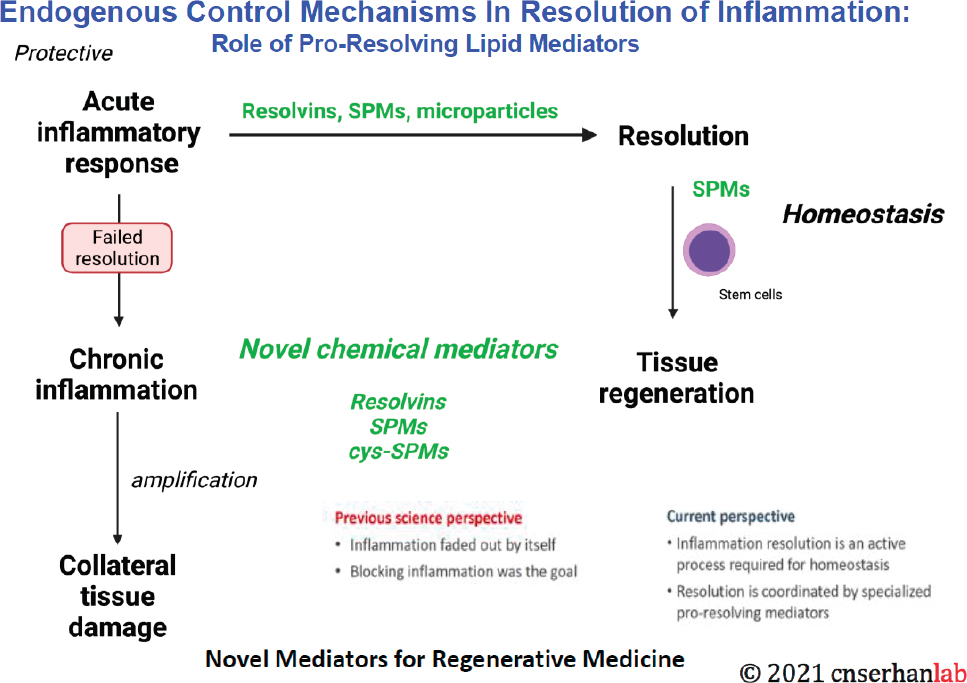
Role of Endogenous Control Mechanisms in Resolving Inflammation
The ideal outcome of an acute inflammatory response is complete resolution of inflammation, and endogenous control mechanisms regulate the resolution process (see Figure 6-2). Interrogating pus from mammals and humans has revealed temporal lipid mediators of class switching. An entire family of specialized pro-resolving lipid mediators functions to stimulate resolution and includes the resolvins, the lipoxins, and the protectins, all of which are derived from essential fatty acids (Serhan and Levy, 2018). The two main functions in resolution consist of (1) the cessation of neutrophil infiltration and (2) phagocytosis of apoptotic polymorphonuclear neutrophils (PMNs) by macrophages, known as efferocytosis, as well as clearance of debris, microbes, and fibrin clots. Investigation of these functions of resolution yielded the structural elucidation of each of the pathways. Serhan and his team introduced resolution indices, which are a quantitative
means of pinpointing where and when the pro-resolving lipid mediators act, he added.
NOTE: SPM = specialized pro-resolving mediator.
SOURCE: Serhan presentation, November 3, 2021.
Eventually, it became evident that stimulating resolution is not equivalent to inhibiting inflammation (i.e., pro-resolution is not anti-inflammation) (Serhan et al., 2015). SPMs activate macrophages to uptake apoptotic PMNs, so they are considered immuno-resolvins that stimulate resolution, Serhan explained. Within the range of potencies of the classic pro-inflammatory mediators, the prostaglandins are situated in the middle. Leukotrienes, which are highly potent pro-inflammatory mediators, are in the picomolar to nanomolar range—as are all pro-resolving mediators. As agonists of resolution, the most potent pro-resolving mediators are often referred to as “stop signals” and include lipoxins, resolvins, protectins, and maresins.
Translational Potential of Specialized Pro-Resolving Mediators
To investigate the action of SPMs in human cells, Serhan and his colleagues used a microfluidic device to simulate human neutrophil swarming and validate “stop” signals. As the first responders to invaders, human
neutrophils “swarm like sharks,” Serhan said. Adding a pro-resolving mediator can stop the neutrophil swarm, providing direct evidence of the action of SPMs on human neutrophils and suggesting that this type of tool could be used to assess immune status (Reategui et al., 2017). Determining each of the biosynthetic pathways and the complete stereochemistry of each of the bioactive mediators that are formed in these cascades from essential fatty acids has provided an opportunity to identify the pro-resolving functions of SPMs (Serhan, 2014). While the primary function is to shorten the resolution interval, other functions of the SPMs include the following:
- Reducing pro-inflammatory cytokines and their counter-regulation
- Reducing eicosanoid storms
- Enhancing PMN clearance and bacterial killing
- Stimulating phagocytosis of apoptotic PMNs (i.e., efferocytosis)
- Increasing removal of inflammatory debris via lymphatics
SPMs are now commercially available and have a range of confirmed functions in animal disease models, including inflammation resolution, tissue protection, infection control, pain reduction, tissue repair, and wound healing. Importantly, these SPMs are log-orders more potent than nonsteroidal anti-inflammatory agents, Serhan added.
Novel Mechanism of Action of Specialized Pro-Resolving Mediators
Most SPMs are produced by two cell types—apoptotic PMNs, which are temporally produced during the trafficking of different cells into the inflammatory milieu, and M2 macrophages—in addition to muscle and adipose tissue, Serhan explained. SPMs work through a novel mechanism of action that limits the magnitude and duration of the acute inflammatory response by reducing both eicosanoid (i.e., bioactive lipid mediator) and cytokine storms. They down-regulate prostaglandins, leukotrienes, and chemokines and cytokines and act in a stereo-selective manner on G-protein–coupled receptors to resolve inflammation. In addition to their actions on the innate immune system, the SPMs and resolvins act on components of the adaptive immune system, including T-cell subsets to regulate cytokine production and produce IL-10 and on B cells to regulate antibody production (Chiurchiu et al., 2016; Serhan et al., 2018). Promising research has shown that SPMs can stimulate stem cells, including periodontal stem cells, neural stem cells, mesenchymal stem cells, and muscle stem cells (Cianci et al., 2016; Dort et al., 2021). For instance, SPMs were shown to accelerate wound closure by stimulating periodontal ligament stem cell migration (Markworth et al., 2020).
Developing Nano Pro-Resolving Medicines
Pro-resolving mediators can be combined with nanoparticles to create nano pro-resolving medicines that mimic endogenous resolution mechanisms, Serhan explained. During the acute inflammatory response, microparticles, which are cell-derived communication vesicles that contain molecular cargo, are released. In the resolution phase of the response, these microvesicles are pro-resolving by carrying intermediates and precursors for resolvins. Serhan and his colleagues mimicked this endogenous resolution phenomenon by creating nanoparticles from human leukocytes and fortifying them with pro-resolving mediators (e.g., resolvin D1) as a therapeutic delivery system (Norling et al., 2011). These enriched nanoparticles have been shown to reduce inflammation in temporomandibular joint (TMJ) inflammation models in humans and to shorten and correct age-related delays in the resolution of acute inflammation in mice (Arnardottir et al., 2014). In the context of periodontal disease, a study in a large-animal model showed that the topical addition of a lipoxin analog, a pro-resolving nanomedicine, reduced inflammation and stimulated bone growth that was quantified by micro-computed tomography (micro-CT) (Van Dyke et al., 2015).
To find evolutionarily conserved elements of regeneration, Serhan and his team turned to planaria, a model organism that is capable of self-regeneration. Studies on the biosynthesis and actions of novel cysteinyl-specialized pro-resolving mediators in planaria head regeneration demonstrate that three pathways are activated in the resolution phase with novel mediators that are peptide–lipid conjugates (Dalli et al., 2014; de la Rosa et al., 2018). Having determined the complete stereochemistry of each of nine pathway molecules and addressed which pathways are activated in the planaria using RNA sequencing, Serhan and his team found that all three activated pathways converge on protein TRAF3 as a key player.3 Moreover, when TRAF3 is silenced in mammalian systems, SPM actions are diminished, and the IL-10 response is lost (Chiang et al., 2021).
Together, this emerging body of evidence indicates that endogenous resolvins and SPMs activate inflammation resolution programs, said Serhan (Serhan, 2017). These mediators are intrinsic controllers of infection, are not immunosuppressant, and can help to lower antibiotic doses. Moreover, they stimulate tissue regeneration, as demonstrated in the planaria study. These data suggest the potential for these pro-resolving mediators in regenerative therapies, he added.
___________________
3 TRAF3 is the tumor necrosis factor receptor-associated factor 3. It is an adaptor protein and considered a “gatekeeper” of certain immune signaling pathways (Chiang et al., 2021).
DISCUSSION
Characteristics of Senescent Cells
Kerr relayed a question about whether senescent stem cells could differentiate, and if so, whether the progeny would carry the senescent phenotype. Kirkland clarified that senescent cells do not divide and exist in a type of dys-differentiated state. They produce factors that impair or impede normal differentiation of cells nearby. Moreover, they can interfere with or accelerate differentiation of different types of cells that are non-senescent. In the context of bone, for example, senescent cells produce factors that can stimulate formation of osteoclasts, which resorb bone, and inhibit formation of osteoblasts, which build new bone. The combination of increasing cells that remove bone and decreasing cells that make new bone appears to contribute to age-related osteoporosis and is being explored in ongoing trials. The same effect holds in progenitors across different systems, he noted. For instance, senescent cells interfere with the differentiated state of adipose cells; fat tissue becomes insulin-resistant as a result. Senescent cells that have a pro-apoptotic SASP also interfere with differentiation of multiple other cell types. Other senescent cells, called “helper” senescent cells, can produce SASP factors (e.g., platelet-derived growth factor-AA) with beneficial effects that can improve wound healing. These types of senescent cells are not targeted by senolytics, but they are targeted in the transgenic p16 depletion mouse models.
Elisseeff added that stem cells can proliferate or they can differentiate. She posited that senescent cells could be considered simply as a differentiation state in a signaling cell. That is, the cells are signaling and communicating with the immune system to create a particular immune environment. She and her colleagues have observed this phenomenon in cartilage, and it reduces tissue production. With this perspective, a stimulus such as an infection in the body could be a signal to not make new tissue. Moreover, senescent cells may be part of the immune rheostat, which determines whether to fight a problem like infection or repair tissue, she said. Kerr added that it would be valuable to further explore the distinction between “good” and “bad” senescence and identify markers to distinguish the two. Elisseeff and her colleagues have used computational techniques to transfer the senescence signature from a transgenic animal to both mouse and human datasets, potentially revealing good-versus-bad senescence cell types and clusters. The results suggest that good types of senescence are associated with angiogenesis-like properties, whereas bad types have properties of fibrosis.
In Vitro Systems to Investigate Senescent Cells
Kirkland was asked if in vitro systems can be used to generate and study senescent cells. He replied that senescent cells can be studied in a range of ways, including in vitro, in vivo, using tissue explants, and so forth. These cells occur across all vertebrate species, with correlates in invertebrate species as well. They are generated in response to multiple kinds of damage signals, including not only aging but also mechanical stress (e.g., in the knee joint with osteoarthritis, or shear stress) and infections of various types. For example, coronavirus can drive cells into senescence, as can bacterial, fungal, and pathogen-associated signals. Intracellular and extracellular damage can also drive senescence. Through mitochondrial communication among cells, senescence can spread through mitochondrial mechanisms as in primitive prokaryotic colonies.
The Senescence Profile and Immunologically Induced Senescence
Kirkland was asked about the long-term effects of the senescence profile on the extracellular matrix, the associated immunological effects, and whether senolytic agents could reverse tissue-level changes. The SASP is complex, varies across different senescent cell types, and evolves over time depending upon the nature of the induced senescence, he explained. Among SASP factors are extracellular matrix proteases, MMPs, and other components that can damage the extracellular matrix. The SASP also contains large amounts of TGF-beta and activin A–related family members that result in fibrosis, as well as various types of micro RNAs that bring in different cell types that can influence the matrix. Furthermore, depending on the senescent cell type, these cells can produce a huge range of chemokines that can attract, activate, and anchor different kinds of immune cells. Senescent cells can do substantial remodeling to the extracellular matrix, he noted. The combination of senolytics and antifibrotics can, therefore, be synergistic in treating disease states associated with fibrosis such as idiopathic pulmonary fibrosis, heart failure with preserved ejection fraction, kidney disease, cirrhosis, sclerosing cholangitis, and a range of other conditions. Clinical trials will soon begin investigating these combinations for treating various kinds of conditions, he added.
Elisseeff commented that aging extracellular matrix can stiffen due not to fibrosis but to loss of proteoglycans with aging. Computational work on senescence has highlighted a role for CCN proteins that are connected to the cytoskeleton.4 Stiffer materials in the extracellular matrix can induce senescence faster; they increase the recruitment of immune cells, such as
___________________
4 For more information about CCN proteins, see Jun and Lau, 2011.
neutrophils and IL-17–producing cells, which also accelerate the onset of senescence. Macrophage cocultures with senescent cells induce more of the receptors and chemokines that communicate with T cells, demonstrating the extent of cell–cell communication between senescent cells and macrophages. The multidirectional communication is complex with T cells influencing the macrophage phenotype. Ongoing mapping efforts seek to clarify the nature and structure of these networks of communication between cell types.
Elisseeff was asked whether IL-17 induces immunosenescence globally or in specific immune cells. Immunosenescence is very different from senescence induction of certain immune phenotypes, she said. She and her colleagues are looking at senescent cell–inducing IL-17 production by various cell types. They have also observed IL-17–producing cells and IL-17–inducing cells that were fibroblasts that were previously healthy, which they refer to as an immunologically induced senescence.
Impact of Mechanics on Response to Biomaterials
Elisseeff was asked about the role of mechanics of biomaterials to shift or otherwise affect the balance between repair and fibrosis—for example, whether immune cell infiltration could be modulated by regulating pore sizes in implanted materials. Indeed, shape, porosity, and mechanics of the materials can influence the response, she said. For instance, stiffer hydro-gels are associated with more fibrosis, greater numbers of neutrophils, and different macrophage phenotypes. These mechanical properties can be modulated, as evidenced in recent papers about the impact of the surface topography of breast implants on fibrosis and serious adverse events in patients. The systemic state of the patient is also relevant and can change over time, Elisseeff added. For example, a patient might receive a cosmetic filler without initial reaction and then have a sudden fibrotic inflammatory response months after injection, which may be due to a systemic change that modulates the local response. Considerations about how the systemic state of an individual may influence the response to injury—with or without a biomaterial—are important from clinical and patient-centered perspectives, she suggested.
Timing of Immune Response Resolution
Serhan was asked about the window of opportunity to capture the resolution of the immune response, processes that took 12–24 hours in the studies he presented. Specifically, he was asked if a short timeframe is common or universal across different tissues or methods of stem cell transplants. Serhan replied that the timing operates differently at each site of
inflammation and in an organ-dependent manner. In the work he presented, the ideal timing was modeled with respect to an inflammatory exudate. With other published animal models, even treatment at the maximum time of the inflammatory response could help trigger endogenous resolution. Thus, the timing and dose of the intervention must be tailored to each system independently, he said. Resolvins, lipoxin analogs, and mimetics have been shown to increase survival in animal models of transplants, Serhan added, which is encouraging and may offer opportunities to synergize with some of the mechanisms discussed by the other presenters.
Applications and Mechanisms of Pro-resolving Mediators
Kerr asked about the mechanisms that the resolvins target within stem cells themselves, and whether they target quiescent cells or replicative stem cells. Serhan and his colleagues have shown that stem cells produce proresolving mediators, as well as prostaglandins and other factors. While other research groups have demonstrated the signal transduction, his group elucidated the receptor themselves. The receptors are present on various stem cells, but after the receptor–ligand interaction, the cellular mechanisms of action are highly cell-type-dependent.
Kerr inquired about the effects of immune response manipulation on stem progenitor cells. Serhan and his lab have demonstrated that periodontal stem cells produce pro-resolving mediators and that these mediators act on neural stem cells. Advances made by other research groups are also promising, including the recent publication demonstrating that resolvin D2 stimulates muscle regeneration, which has the potential for treating Duchenne muscular dystrophy (Dort et al., 2021).
Serhan was also asked what happens if neutrophils or macrophages are absent or blocked, and whether it would be effective to add mediators of their function. He replied that it is unlikely that the complex role of the macrophage—particularly the M2 macrophage—or the neutrophil could be substituted by a single mediator that they produce or that acts on them. However, if they are absent from the system, other targets have been elucidated for pro-resolving mediators, and their receptors appear in other tissues, representing opportunities for future study.
Manipulation of Immune Responses and Autoimmunity Issues
Kerr asked about any concerns or evidence that manipulating immune responses might generate autoimmunity issues. Elisseeff replied that the corollary of that question is whether manipulating immune responses can be used to treat autoimmune disease in new ways. The IL-17 signature, for
instance, is associated with certain autoimmune diseases such as psoriatic arthritis. Resolving a wound in various ways could also help determine the signatures of autoimmunity. There are potential benefits of manipulating immune responses as well as dangers, she said. Serhan added that evidence is emerging about the effects of resolvins on autoimmunity for osteoarthritis and rheumatoid arthritis, for example, in which the administration of resolvins appears to result in a diminished inflammatory response and improved cartilage production and function. He suggested that there are many opportunities to consider the appropriate indications and disease scenarios to deliver pro-resolving mediators, which is why his group has used planaria to investigate highly conserved components of tissue regeneration that intersect with the actions of pro-resolving mediators.
This page intentionally left blank.


